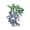
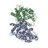
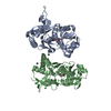
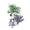
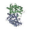
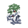
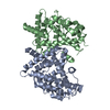
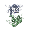
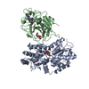
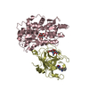
Entry Database : PDB / ID : 6qf7Title Crystal structures of the recombinant beta-Factor XIIa protease with bound Thr-Arg and Pro-Arg substrate mimetics Coagulation factor XII Maltodextrin-binding protein Keywords / / / / Function / homology Function Domain/homology Component
/ / / / / / / / / / / / / / / / / / / / / / / / / / / / / / / / / / / / / / / / / / / / / / / / / / / / / / / / / / / / / / / / / / / / / / / / / / / / / / / / / / / / / / / / / / / / / Biological species Escherichia coli (E. coli)Homo sapiens (human)Method / / / Resolution : 4 Å Authors Pathak, M. / Mannal, R. / Li, C. / Bubacarr, G.K. / Badraldin, K.H. / Belviso, B.D. / Camila, R.B. / Dreveny, I. / Fischer, P.M. / Dekker, L.V. ...Pathak, M. / Mannal, R. / Li, C. / Bubacarr, G.K. / Badraldin, K.H. / Belviso, B.D. / Camila, R.B. / Dreveny, I. / Fischer, P.M. / Dekker, L.V. / Oliva, M.L.V. / Emsley, J. Funding support Organization Grant number Country British Heart Foundation RG/12/9/29775
Journal : Acta Crystallogr D Struct Biol / Year : 2019Title : Crystal structures of the recombinant beta-factor XIIa protease with bound Thr-Arg and Pro-Arg substrate mimetics.Authors : Pathak, M. / Manna, R. / Li, C. / Kaira, B.G. / Hamad, B.K. / Belviso, B.D. / Bonturi, C.R. / Dreveny, I. / Fischer, P.M. / Dekker, L.V. / Oliva, M.L.V. / Emsley, J. History Deposition Jan 9, 2019 Deposition site / Processing site Revision 1.0 Jun 5, 2019 Provider / Type Revision 1.1 Jun 26, 2019 Group / Database referencesCategory citation / citation_author ... citation / citation_author / database_PDB_rev / database_PDB_rev_record / pdbx_database_proc Item _citation.country / _citation.journal_abbrev ... _citation.country / _citation.journal_abbrev / _citation.journal_id_ASTM / _citation.journal_id_ISSN / _citation.journal_volume / _citation.page_first / _citation.page_last / _citation.pdbx_database_id_PubMed / _citation.title Revision 1.2 Jul 31, 2019 Group / Structure summary / Category Revision 2.0 Jul 29, 2020 Group Advisory / Atomic model ... Advisory / Atomic model / Data collection / Derived calculations / Non-polymer description / Structure summary Category atom_site / chem_comp ... atom_site / chem_comp / database_PDB_caveat / entity / entity_name_com / pdbx_branch_scheme / pdbx_chem_comp_identifier / pdbx_entity_branch / pdbx_entity_branch_descriptor / pdbx_entity_branch_link / pdbx_entity_branch_list / pdbx_entity_nonpoly / pdbx_molecule / pdbx_molecule_features / pdbx_nonpoly_scheme / pdbx_struct_assembly_gen / pdbx_unobs_or_zero_occ_atoms / pdbx_validate_chiral / pdbx_validate_close_contact / struct_asym / struct_conn / struct_site / struct_site_gen Item _atom_site.B_iso_or_equiv / _atom_site.Cartn_x ... _atom_site.B_iso_or_equiv / _atom_site.Cartn_x / _atom_site.Cartn_y / _atom_site.Cartn_z / _atom_site.auth_asym_id / _atom_site.auth_atom_id / _atom_site.auth_comp_id / _atom_site.auth_seq_id / _atom_site.label_asym_id / _atom_site.label_atom_id / _atom_site.label_comp_id / _atom_site.label_entity_id / _atom_site.occupancy / _atom_site.type_symbol / _chem_comp.formula / _chem_comp.formula_weight / _chem_comp.id / _chem_comp.mon_nstd_flag / _chem_comp.name / _chem_comp.type / _database_PDB_caveat.text / _pdbx_struct_assembly_gen.asym_id_list / _pdbx_unobs_or_zero_occ_atoms.auth_asym_id / _pdbx_unobs_or_zero_occ_atoms.auth_seq_id / _pdbx_unobs_or_zero_occ_atoms.label_asym_id / _pdbx_validate_chiral.auth_asym_id / _pdbx_validate_chiral.auth_seq_id / _pdbx_validate_chiral.details / _pdbx_validate_close_contact.auth_asym_id_2 / _pdbx_validate_close_contact.auth_seq_id_2 Description / Provider / Type Revision 2.1 Nov 6, 2024 Group Advisory / Data collection ... Advisory / Data collection / Database references / Structure summary Category chem_comp / chem_comp_atom ... chem_comp / chem_comp_atom / chem_comp_bond / database_2 / pdbx_entry_details / pdbx_modification_feature / pdbx_unobs_or_zero_occ_atoms Item / _database_2.pdbx_DOI / _database_2.pdbx_database_accession
Show all Show less
 Movie
Movie Controller

 Yorodumi
 Open data
 Basic information
 Components
 Keywords
 Function and homology information
 Homo sapiens (human)
 X-RAY DIFFRACTION /
  Authors
 United Kingdom, 1items
  Citation
 Structure visualization
 Downloads & links
 Other downloads
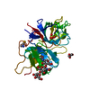


 PDBj
PDBj


 Assembly
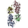


 Type: D-saccharide, beta linking / Mass: 221.208 Da / Num. of mol.: 1
 Type: peptide-like, Peptide-like / Class: Inhibitor / Mass: 453.986 Da / Num. of mol.: 2 / Source method: obtained synthetically / Formula: C21H34ClN6O3 / References: D-Phe-Pro-Arg-CH2Cl
 Sample preparation
 Processing