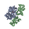
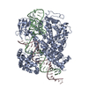
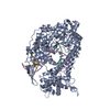
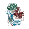
[English] 日本語
Yorodumi
- PDB-3u93: Crystal structure of the GluK3 ligand binding domain complex with... -

+
Open data


ID or keywords:

Loading...

-
Basic information

Entry
Database: PDB / ID: 3u93
TitleCrystal structure of the GluK3 ligand binding domain complex with glutamate and zinc: P2221 form
ComponentsGlutamate receptor, ionotropic kainate 3
KeywordsMEMBRANE PROTEIN / ION CHANNEL
Function / homology
Function and homology information


Presynaptic function of Kainate receptors / adenylate cyclase inhibiting G protein-coupled glutamate receptor activity / G protein-coupled glutamate receptor signaling pathway / kainate selective glutamate receptor complex / Activation of Ca-permeable Kainate Receptor / negative regulation of synaptic transmission, glutamatergic / glutamate receptor signaling pathway / glutamate receptor activity / kainate selective glutamate receptor activity / glutamate-gated receptor activity ...Presynaptic function of Kainate receptors / adenylate cyclase inhibiting G protein-coupled glutamate receptor activity / G protein-coupled glutamate receptor signaling pathway / kainate selective glutamate receptor complex / Activation of Ca-permeable Kainate Receptor / negative regulation of synaptic transmission, glutamatergic / glutamate receptor signaling pathway / glutamate receptor activity / kainate selective glutamate receptor activity / glutamate-gated receptor activity / ligand-gated monoatomic ion channel activity involved in regulation of presynaptic membrane potential / dendrite cytoplasm / synaptic transmission, glutamatergic / transmitter-gated monoatomic ion channel activity involved in regulation of postsynaptic membrane potential / regulation of membrane potential / postsynaptic density membrane / modulation of chemical synaptic transmission / terminal bouton / presynaptic membrane / chemical synaptic transmission / perikaryon / axon / glutamatergic synapse / dendrite / plasma membrane
Similarity search - Function
Ionotropic glutamate receptor, metazoa / Ligated ion channel L-glutamate- and glycine-binding site / : / Ligand-gated ion channel / Ionotropic glutamate receptor, L-glutamate and glycine-binding domain / Ligated ion channel L-glutamate- and glycine-binding site / Ionotropic glutamate receptor / Eukaryotic homologues of bacterial periplasmic substrate binding proteins. / Receptor, ligand binding region / Receptor family ligand binding region ...Ionotropic glutamate receptor, metazoa / Ligated ion channel L-glutamate- and glycine-binding site / : / Ligand-gated ion channel / Ionotropic glutamate receptor, L-glutamate and glycine-binding domain / Ligated ion channel L-glutamate- and glycine-binding site / Ionotropic glutamate receptor / Eukaryotic homologues of bacterial periplasmic substrate binding proteins. / Receptor, ligand binding region / Receptor family ligand binding region / Periplasmic binding protein-like II / Periplasmic binding protein-like I / D-Maltodextrin-Binding Protein; domain 2 / 3-Layer(aba) Sandwich / Alpha Beta
Similarity search - Domain/homology
GLUTAMIC ACID / Glutamate receptor ionotropic, kainate 3
Similarity search - Component
Biological speciesRattus norvegicus (Norway rat)
MethodX-RAY DIFFRACTION / SYNCHROTRON / MOLECULAR REPLACEMENT / Resolution: 1.881 Å
AuthorsKumar, J. / Mayer, M.L.
CitationJournal: Neuron / Year: 2012
Title: Zinc Potentiates GluK3 Glutamate Receptor Function by Stabilizing the Ligand Binding Domain Dimer Interface.
Authors: Veran, J. / Kumar, J. / Pinheiro, P.S. / Athane, A. / Mayer, M.L. / Perrais, D. / Mulle, C.
History
DepositionOct 17, 2011Deposition site: RCSB / Processing site: RCSB
Revision 1.0Sep 26, 2012Provider: repository / Type: Initial release
Revision 1.1Dec 19, 2012Group: Database references
Revision 1.2Aug 12, 2015Group: Other
Revision 1.3Aug 9, 2017Group: Refinement description / Source and taxonomy / Category: entity_src_gen / software
Revision 1.4Sep 13, 2023Group: Data collection / Database references ...Data collection / Database references / Derived calculations / Refinement description
Category: chem_comp_atom / chem_comp_bond ...chem_comp_atom / chem_comp_bond / database_2 / pdbx_initial_refinement_model / pdbx_struct_conn_angle / struct_conn / struct_ref_seq_dif / struct_site
Item: _database_2.pdbx_DOI / _database_2.pdbx_database_accession ..._database_2.pdbx_DOI / _database_2.pdbx_database_accession / _pdbx_struct_conn_angle.ptnr1_auth_asym_id / _pdbx_struct_conn_angle.ptnr1_auth_comp_id / _pdbx_struct_conn_angle.ptnr1_auth_seq_id / _pdbx_struct_conn_angle.ptnr1_label_asym_id / _pdbx_struct_conn_angle.ptnr1_label_atom_id / _pdbx_struct_conn_angle.ptnr1_label_comp_id / _pdbx_struct_conn_angle.ptnr1_label_seq_id / _pdbx_struct_conn_angle.ptnr2_auth_asym_id / _pdbx_struct_conn_angle.ptnr2_label_asym_id / _pdbx_struct_conn_angle.ptnr3_auth_asym_id / _pdbx_struct_conn_angle.ptnr3_auth_comp_id / _pdbx_struct_conn_angle.ptnr3_auth_seq_id / _pdbx_struct_conn_angle.ptnr3_label_asym_id / _pdbx_struct_conn_angle.ptnr3_label_atom_id / _pdbx_struct_conn_angle.ptnr3_label_comp_id / _pdbx_struct_conn_angle.ptnr3_label_seq_id / _pdbx_struct_conn_angle.value / _struct_conn.pdbx_dist_value / _struct_conn.ptnr1_auth_asym_id / _struct_conn.ptnr1_auth_comp_id / _struct_conn.ptnr1_auth_seq_id / _struct_conn.ptnr1_label_asym_id / _struct_conn.ptnr1_label_atom_id / _struct_conn.ptnr1_label_comp_id / _struct_conn.ptnr1_label_seq_id / _struct_conn.ptnr2_auth_asym_id / _struct_conn.ptnr2_auth_comp_id / _struct_conn.ptnr2_auth_seq_id / _struct_conn.ptnr2_label_asym_id / _struct_conn.ptnr2_label_atom_id / _struct_conn.ptnr2_label_comp_id / _struct_ref_seq_dif.details / _struct_site.pdbx_auth_asym_id / _struct_site.pdbx_auth_comp_id / _struct_site.pdbx_auth_seq_id

-
Structure visualization

Structure viewerMolecule:
MolmilJmol/JSmol

Downloads & links

-
Assembly

Deposited unit
A: Glutamate receptor, ionotropic kainate 3
B: Glutamate receptor, ionotropic kainate 3
hetero molecules


Theoretical massNumber of molelcules
Total (without water)58,79911
Polymers58,1072
Non-polymers6929
Water3,909217
1
A: Glutamate receptor, ionotropic kainate 3
B: Glutamate receptor, ionotropic kainate 3
hetero molecules

A: Glutamate receptor, ionotropic kainate 3
B: Glutamate receptor, ionotropic kainate 3
hetero molecules


Theoretical massNumber of molelcules
Total (without water)117,59822
Polymers116,2134
Non-polymers1,38418
Water724
TypeNameSymmetry operationNumber
identity operation1_555x,y,z1
crystal symmetry operation4_565x,-y+1,-z1
Buried area4800 Å2
ΔGint-320 kcal/mol
Surface area46620 Å2
MethodPISA
Unit cell
Length a, b, c (Å)130.401, 55.457, 88.861
Angle α, β, γ (deg.)90.00, 90.00, 90.00
Int Tables number17
Space group name H-MP2221
DetailsTHE OBSERVED BIOLOGICAL UNIT DOES NOT OCCUR FOR THE FULL LENGTH PROTEIN, THEREFORE, IT IS NOT BIOLOGICALLY RELEVANT

-
Components

#1: Protein Glutamate receptor, ionotropic kainate 3 / GluK3


Mass: 29053.371 Da / Num. of mol.: 2 / Fragment: unp residues 433-546 and 669-807
Source method: isolated from a genetically manipulated source
Source: (gene. exp.) Rattus norvegicus (Norway rat) / Gene: GRIK3 / Plasmid: PET22 modified / Production host: Escherichia coli (E. coli) / Strain (production host): ORIGAMI B(DE3) / References: UniProt: P42264
#2: Chemical ChemComp-GLU / GLUTAMIC ACID


Type: L-peptide linking / Mass: 147.129 Da / Num. of mol.: 2 / Source method: obtained synthetically / Formula: C5H9NO4
#3: Chemical
ChemComp-ZN / ZINC ION


Mass: 65.409 Da / Num. of mol.: 5 / Source method: obtained synthetically / Formula: Zn
#4: Chemical ChemComp-CL / CHLORIDE ION


Mass: 35.453 Da / Num. of mol.: 2 / Source method: obtained synthetically / Formula: Cl
#5: Water ChemComp-HOH / water


Mass: 18.015 Da / Num. of mol.: 217 / Source method: isolated from a natural source / Formula: H2O
Sequence detailsTHE PROTEIN CRYSTALLIZED IS THE EXTRACELLULAR LIGAND BINDING DOMAIN OF GLUK3. TRANSMEMBRANE REGIONS ...THE PROTEIN CRYSTALLIZED IS THE EXTRACELLULAR LIGAND BINDING DOMAIN OF GLUK3. TRANSMEMBRANE REGIONS WERE GENETICALLY REMOVED AND REPLACED WITH A GLY-THR LINKER THEREFORE, THE SEQUENCE MATCHES DISCONTINUOUSLY WITH THE REFERENCE DATABASE (433-546 AND 669-807)

-
Experimental details

-
Experiment

ExperimentMethod: X-RAY DIFFRACTION / Number of used crystals: 1

-
Sample preparation

CrystalDensity Matthews: 2.76 Å3/Da / Density % sol: 55.51 %
Crystal growTemperature: 293 K / Method: vapor diffusion, hanging drop / pH: 8.4
Details: 14% PEG 3350, 0.1 M BisTrisPropane, 0.2 M Na2SO4, 5 mM glutamate, 5 mM ZnAcetate, pH 8.4, VAPOR DIFFUSION, HANGING DROP, temperature 293K

-
Data collection

DiffractionMean temperature: 100 K
Diffraction sourceSource: SYNCHROTRON / Site: APS / Beamline: 22-ID / Wavelength: 1 Å
DetectorType: MARMOSAIC 300 mm CCD / Detector: CCD / Date: Apr 7, 2011
RadiationMonochromator: Si(111) / Protocol: SINGLE WAVELENGTH / Monochromatic (M) / Laue (L): M / Scattering type: x-ray
Radiation wavelengthWavelength: 1 Å / Relative weight: 1
ReflectionResolution: 1.88→40 Å / Num. all: 53068 / Num. obs: 53068 / % possible obs: 98.4 % / Observed criterion σ(F): 0 / Observed criterion σ(I): 0 / Redundancy: 5.6 % / Biso Wilson estimate: 31.1 Å2 / Rmerge(I) obs: 0.049 / Net I/σ(I): 23.8
Reflection shellResolution: 1.88→1.91 Å / Redundancy: 5.5 % / Rmerge(I) obs: 0.473 / Mean I/σ(I) obs: 2.8 / % possible all: 95.2

-
Processing

Software
NameVersionClassification
HKL-2000data collection
PHASERphasing
PHENIX(phenix.refine: 1.7_650)refinement
HKL-2000data reduction
HKL-2000data scaling
RefinementMethod to determine structure: MOLECULAR REPLACEMENT
Starting model: 1S50
Resolution: 1.881→39.046 Å / SU ML: 0.25 / Cross valid method: THROUGHOUT / σ(F): 0 / Phase error: 26.88 / Stereochemistry target values: ML
RfactorNum. reflection% reflectionSelection details
Rfree0.2483 2677 5.13 %RANDOM
Rwork0.2142 ---
all0.2159 52177 --
obs0.2159 52177 98.13 %-
Solvent computationShrinkage radii: 0.89 Å / VDW probe radii: 1 Å / Solvent model: FLAT BULK SOLVENT MODEL / Bsol: 40.727 Å2 / ksol: 0.398 e/Å3
Displacement parameters
Baniso -1Baniso -2Baniso -3
1--10.8947 Å2-0 Å20 Å2
2--1.8151 Å20 Å2
3---9.0796 Å2
Refinement stepCycle: LAST / Resolution: 1.881→39.046 Å
ProteinNucleic acidLigandSolventTotal
Num. atoms4005 0 27 217 4249
Refine LS restraints
Refine-IDTypeDev idealNumber
X-RAY DIFFRACTIONf_bond_d0.014123
X-RAY DIFFRACTIONf_angle_d0.9185544
X-RAY DIFFRACTIONf_dihedral_angle_d13.631554
X-RAY DIFFRACTIONf_chiral_restr0.054614
X-RAY DIFFRACTIONf_plane_restr0.004695
LS refinement shell
Resolution (Å)Rfactor RfreeNum. reflection RfreeRfactor RworkNum. reflection RworkRefine-ID% reflection obs (%)
1.8813-1.91550.3491210.30362377X-RAY DIFFRACTION90
1.9155-1.95240.39761380.30152550X-RAY DIFFRACTION99
1.9524-1.99220.29511440.27492544X-RAY DIFFRACTION96
1.9922-2.03550.29791690.26092527X-RAY DIFFRACTION98
2.0355-2.08290.26251440.24242535X-RAY DIFFRACTION97
2.0829-2.1350.29211470.22292558X-RAY DIFFRACTION97
2.135-2.19270.24681280.22552603X-RAY DIFFRACTION99
2.1927-2.25720.26951540.22212537X-RAY DIFFRACTION98
2.2572-2.330.2441220.22872636X-RAY DIFFRACTION98
2.33-2.41330.29081630.23462581X-RAY DIFFRACTION99
2.4133-2.50990.27911210.24542630X-RAY DIFFRACTION100
2.5099-2.62410.29471420.23982649X-RAY DIFFRACTION99
2.6241-2.76240.30951230.2452624X-RAY DIFFRACTION99
2.7624-2.93550.26131420.24742627X-RAY DIFFRACTION99
2.9355-3.1620.25681360.23342672X-RAY DIFFRACTION100
3.162-3.480.27561410.22762659X-RAY DIFFRACTION100
3.48-3.98320.2241640.20132674X-RAY DIFFRACTION100
3.9832-5.01670.19391420.15842722X-RAY DIFFRACTION99
5.0167-39.05430.21291360.19312795X-RAY DIFFRACTION97
Refinement TLS params.

Method: refined / Refine-ID: X-RAY DIFFRACTION

IDL112)L122)L132)L222)L232)L332)S11 (Å °)S12 (Å °)S13 (Å °)S21 (Å °)S22 (Å °)S23 (Å °)S31 (Å °)S32 (Å °)S33 (Å °)T112)T122)T132)T222)T232)T332)Origin x (Å)Origin y (Å)Origin z (Å)
10.04410.06690.02260.1157-0.02580.2450.0430.0075-0.0889-0.07810.0825-0.04270.129-0.0701-0.05270.27-0.11860.0930.2003-0.02520.931613.04595.888211.1856
20.4446-0.39280.02270.3982-0.03880.1090.0891-0.169-0.2843-0.03770.0740.4938-0.0039-0.02310.06370.2202-0.04810.17880.2167-0.10480.566222.164819.109617.516
30.0695-0.0455-0.01010.02890.01090.0269-0.0393-0.15050.02830.03840.05790.01450.00110.0294-0.01280.3156-0.04720.24920.4949-0.15690.447921.720129.370528.7015
40.1761-0.02530.22330.0013-0.02850.29210.1504-0.0989-0.04510.0430.07690.2006-0.0904-0.0361-0.12560.268-0.02810.18140.2853-0.07050.562914.163326.822616.9126
50.0245-0.011-0.00320.02070.00770.0118-0.02090.0617-0.0582-0.0178-0.03610.0228-0.0325-0.05840.01980.25230.0531-0.01460.3445-0.20620.402329.572514.8683-0.348
60.01710.06050.0030.21990.00870.00160.01620.1591-0.24220.06290.01990.1646-0.0325-0.0434-0.02380.2348-0.0009-0.00240.3517-0.11690.536711.466522.55511.6884
70.1579-0.0664-0.00990.1501-0.05860.0316-0.1144-0.0275-0.01970.0115-0.0283-0.07590.11740.19230.1310.265-0.02440.02820.2599-0.0360.199458.499835.220811.2594
80.8241-0.0413-0.04120.1801-0.25450.3885-0.11790.13280.37910.1105-0.1773-0.005-0.2126-0.01290.18440.3769-0.0591-0.09930.2778-0.08510.342553.570249.134310.3543
90.4277-0.0373-0.09430.11640.05110.033-0.2069-0.43880.08560.09710.13580.04980.0117-0.10930.01440.33820.0786-0.00680.4217-0.17670.218542.657243.612324.6691
100.1292-0.02190.00350.170.06980.426-0.2505-0.38510.18150.04970.04140.0292-0.0876-0.2547-0.08230.26760.1438-0.06210.5115-0.40260.120147.051545.162831.0949
110.310.00280.04490.2838-0.24250.6307-0.15770.03260.03910.0235-0.16590.05160.1-0.20180.27480.2813-0.07440.02860.3687-0.10820.243842.112633.03247.9196
120.04870.0186-0.01410.0249-0.00140.00640.0401-0.0406-0.00740.0023-0.0211-0.01380.0014-0.0042-0.01420.3227-0.15720.04990.3734-0.01440.240448.262625.669324.6342
Refinement TLS group
IDRefine-IDRefine TLS-IDSelection detailsAuth asym-IDAuth seq-ID
1X-RAY DIFFRACTION1( CHAIN A AND RESID 3:31 )A3 - 31
2X-RAY DIFFRACTION2( CHAIN A AND RESID 32:139 )A32 - 139
3X-RAY DIFFRACTION3( CHAIN A AND RESID 140:173 )A140 - 173
4X-RAY DIFFRACTION4( CHAIN A AND RESID 174:223 )A174 - 223
5X-RAY DIFFRACTION5( CHAIN A AND RESID 224:240 )A224 - 240
6X-RAY DIFFRACTION6( CHAIN A AND ( RESID 241:251 OR RESID 254:256 ) )A241 - 251
7X-RAY DIFFRACTION6( CHAIN A AND ( RESID 241:251 OR RESID 254:256 ) )A254 - 256
8X-RAY DIFFRACTION7( CHAIN B AND RESID 3:42 )B3 - 42
9X-RAY DIFFRACTION8( CHAIN B AND RESID 43:83 )B43 - 83
10X-RAY DIFFRACTION9( CHAIN B AND RESID 84:127 )B84 - 127
11X-RAY DIFFRACTION10( CHAIN B AND RESID 128:223 )B128 - 223
12X-RAY DIFFRACTION11( CHAIN B AND RESID 224:249 )B224 - 249
13X-RAY DIFFRACTION12( CHAIN B AND ( RESID 250:251 OR RESID 255:255 ) )B250 - 251
14X-RAY DIFFRACTION12( CHAIN B AND ( RESID 250:251 OR RESID 255:255 ) )B255

+
About Yorodumi

-
News

-
Feb 9, 2022. New format data for meta-information of EMDB entries

New format data for meta-information of EMDB entries

  • Version 3 of the EMDB header file is now the official format.
  • The previous official version 1.9 will be removed from the archive.

Related info.:EMDB header

External links:wwPDB to switch to version 3 of the EMDB data model

-
Aug 12, 2020. Covid-19 info

Covid-19 info

URL: https://pdbjlvh1.pdbj.org/emnavi/covid19.php

New page: Covid-19 featured information page in EM Navigator.

Related info.:Covid-19 info / Mar 5, 2020. Novel coronavirus structure data

+
Mar 5, 2020. Novel coronavirus structure data

Novel coronavirus structure data

Related info.:Yorodumi Speices / Aug 12, 2020. Covid-19 info

External links:COVID-19 featured content - PDBj / Molecule of the Month (242):Coronavirus Proteases

+
Jan 31, 2019. EMDB accession codes are about to change! (news from PDBe EMDB page)

EMDB accession codes are about to change! (news from PDBe EMDB page)

  • The allocation of 4 digits for EMDB accession codes will soon come to an end. Whilst these codes will remain in use, new EMDB accession codes will include an additional digit and will expand incrementally as the available range of codes is exhausted. The current 4-digit format prefixed with “EMD-” (i.e. EMD-XXXX) will advance to a 5-digit format (i.e. EMD-XXXXX), and so on. It is currently estimated that the 4-digit codes will be depleted around Spring 2019, at which point the 5-digit format will come into force.
  • The EM Navigator/Yorodumi systems omit the EMD- prefix.

Related info.:Q: What is EMD? / ID/Accession-code notation in Yorodumi/EM Navigator

External links:EMDB Accession Codes are Changing Soon! / Contact to PDBj

+
Jul 12, 2017. Major update of PDB

Major update of PDB

  • wwPDB released updated PDB data conforming to the new PDBx/mmCIF dictionary.
  • This is a major update changing the version number from 4 to 5, and with Remediation, in which all the entries are updated.
  • In this update, many items about electron microscopy experimental information are reorganized (e.g. em_software).
  • Now, EM Navigator and Yorodumi are based on the updated data.

External links:wwPDB Remediation / Enriched Model Files Conforming to OneDep Data Standards Now Available in the PDB FTP Archive

-
Yorodumi

Thousand views of thousand structures

  • Yorodumi is a browser for structure data from EMDB, PDB, SASBDB, etc.
  • This page is also the successor to EM Navigator detail page, and also detail information page/front-end page for Omokage search.
  • The word "yorodu" (or yorozu) is an old Japanese word meaning "ten thousand". "mi" (miru) is to see.

Related info.:EMDB / PDB / SASBDB / Comparison of 3 databanks / Yorodumi Search / Aug 31, 2016. New EM Navigator & Yorodumi / Yorodumi Papers / Jmol/JSmol / Function and homology information / Changes in new EM Navigator and Yorodumi

Read more