Mass: 18.015 Da / Num. of mol.: 581 / Source method: isolated from a natural source / Formula: H2O
-
Details
Has protein modification
Y
Sequence details
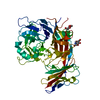
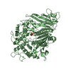
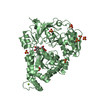
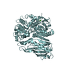
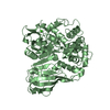
THE PROTEIN CRYSTALLIZED IS THE EXTRACELLULAR LIGAND BINDING DOMAIN OF GLUK3. TRANSMEMBRANE REGIONS ...THE PROTEIN CRYSTALLIZED IS THE EXTRACELLULAR LIGAND BINDING DOMAIN OF GLUK3. TRANSMEMBRANE REGIONS WERE GENETICALLY REMOVED AND REPLACED WITH A GLY-THR LINKER (RESIDUES 119 AND 120 OF THE STRUCTURE). THEREFORE, THE SEQUENCE MATCHES DISCONTINUOUSLY WITH THE REFERENCE DATABASE (432-546, 669-806)
-
Experimental details
-
Experiment
Experiment
Method: X-RAY DIFFRACTION / Number of used crystals: 1
-
Sample preparation
Crystal
Density Matthews: 2.42 Å3/Da / Density % sol: 49.13 %
Crystal grow
Temperature: 293 K / Method: vapor diffusion, hanging drop / pH: 8.2 Details: 1.8 M sodium/potassium phosphate, pH 8.2, VAPOR DIFFUSION, HANGING DROP, temperature 293K
Resolution: 1.6→27.897 Å / SU ML: 0.36 / Isotropic thermal model: Restrained / Cross valid method: THROUGHOUT / σ(F): 0 / Phase error: 16.82 / Stereochemistry target values: ML Details: Residues 1-2 (GP) and residues 254-256 (GSG) in both chains were not located in the electron density map.
Rfactor
Num. reflection
% reflection
Selection details
Rfree
0.1837
3664
5.06 %
Random
Rwork
0.1352
-
-
-
obs
0.1377
72446
99.75 %
-
all
-
72508
-
-
Solvent computation
Shrinkage radii: 1.01 Å / VDW probe radii: 1.1 Å / Solvent model: FLAT BULK SOLVENT MODEL / Bsol: 38.722 Å2 / ksol: 0.461 e/Å3
Displacement parameters
Biso mean: 20.627 Å2
Baniso -1
Baniso -2
Baniso -3
1-
0.5033 Å2
0 Å2
-0 Å2
2-
-
0.5033 Å2
-0 Å2
3-
-
-
-1.0066 Å2
Refinement step
Cycle: LAST / Resolution: 1.6→27.897 Å
Protein
Nucleic acid
Ligand
Solvent
Total
Num. atoms
4032
0
48
581
4661
Refine LS restraints
Refine-ID
Type
Dev ideal
Number
X-RAY DIFFRACTION
f_bond_d
0.013
4229
X-RAY DIFFRACTION
f_angle_d
1.29
5705
X-RAY DIFFRACTION
f_dihedral_angle_d
13.848
1612
X-RAY DIFFRACTION
f_chiral_restr
0.079
631
X-RAY DIFFRACTION
f_plane_restr
0.006
721
LS refinement shell
Refine-ID: X-RAY DIFFRACTION
Resolution (Å)
Rfactor Rfree
Num. reflection Rfree
Rfactor Rwork
Num. reflection Rwork
Num. reflection obs
% reflection obs (%)
1.6-1.6211
0.2713
132
0.1828
2603
2603
99
1.6211-1.6433
0.2336
128
0.1647
2615
2615
99
1.6433-1.6668
0.2562
139
0.1674
2655
2655
99
1.6668-1.6916
0.2343
140
0.163
2631
2631
99
1.6916-1.7181
0.2629
136
0.1577
2579
2579
99
1.7181-1.7462
0.2442
145
0.1413
2672
2672
99
1.7462-1.7763
0.2071
134
0.1285
2652
2652
100
1.7763-1.8086
0.2156
145
0.1339
2620
2620
100
1.8086-1.8434
0.2006
148
0.1275
2617
2617
100
1.8434-1.881
0.1795
117
0.1293
2723
2723
100
1.881-1.9219
0.1832
146
0.1231
2598
2599
100
1.9219-1.9666
0.198
145
0.12
2665
2665
100
1.9666-2.0158
0.1686
140
0.1122
2623
2623
100
2.0158-2.0703
0.1695
155
0.1141
2662
2662
100
2.0703-2.1312
0.1699
151
0.1144
2615
2615
100
2.1312-2.1999
0.1857
136
0.1085
2667
2667
100
2.1999-2.2785
0.1671
147
0.1117
2638
2638
100
2.2785-2.3697
0.1607
141
0.1158
2660
2660
100
2.3697-2.4775
0.1683
133
0.1253
2639
2639
100
2.4775-2.608
0.1959
132
0.1301
2692
2692
100
2.608-2.7713
0.1806
157
0.1283
2623
2623
100
2.7713-2.985
0.1715
141
0.133
2659
2659
100
2.985-3.285
0.1722
135
0.1399
2649
2649
100
3.285-3.7594
0.1615
155
0.1426
2669
2669
100
3.7594-4.7326
0.1687
150
0.1243
2656
2657
100
4.7326-27.9008
0.2032
136
0.1886
2700
2707
99
+
About Yorodumi
-
News
-
Feb 9, 2022. New format data for meta-information of EMDB entries
New format data for meta-information of EMDB entries
Version 3 of the EMDB header file is now the official format.
The previous official version 1.9 will be removed from the archive.
In the structure databanks used in Yorodumi, some data are registered as the other names, "COVID-19 virus" and "2019-nCoV". Here are the details of the virus and the list of structure data.
Jan 31, 2019. EMDB accession codes are about to change! (news from PDBe EMDB page)
EMDB accession codes are about to change! (news from PDBe EMDB page)
The allocation of 4 digits for EMDB accession codes will soon come to an end. Whilst these codes will remain in use, new EMDB accession codes will include an additional digit and will expand incrementally as the available range of codes is exhausted. The current 4-digit format prefixed with “EMD-” (i.e. EMD-XXXX) will advance to a 5-digit format (i.e. EMD-XXXXX), and so on. It is currently estimated that the 4-digit codes will be depleted around Spring 2019, at which point the 5-digit format will come into force.
The EM Navigator/Yorodumi systems omit the EMD- prefix.
Related info.:Q: What is EMD? / ID/Accession-code notation in Yorodumi/EM Navigator
Yorodumi is a browser for structure data from EMDB, PDB, SASBDB, etc.
This page is also the successor to EM Navigator detail page, and also detail information page/front-end page for Omokage search.
The word "yorodu" (or yorozu) is an old Japanese word meaning "ten thousand". "mi" (miru) is to see.
Related info.:EMDB / PDB / SASBDB / Comparison of 3 databanks / Yorodumi Search / Aug 31, 2016. New EM Navigator & Yorodumi / Yorodumi Papers / Jmol/JSmol / Function and homology information / Changes in new EM Navigator and Yorodumi
 Movie
Movie Controller

 Yorodumi
 Open data
 Basic information
 Components
 Keywords
 Function and homology information
 X-RAY DIFFRACTION /
  Authors
 Citation
 Structure visualization
 Downloads & links
 Other downloads


 PDBj
PDBj


 Assembly


 Type: L-peptide linking / Mass: 147.129 Da / Num. of mol.: 2 / Source method: obtained synthetically / Formula: C5H9NO4
 Mass: 92.094 Da / Num. of mol.: 1 / Source method: obtained synthetically / Formula: C3H8O3
 Mass: 35.453 Da / Num. of mol.: 4 / Source method: obtained synthetically / Formula: Cl
 Mass: 94.971 Da / Num. of mol.: 3 / Source method: obtained synthetically / Formula: PO4
 Mass: 22.990 Da / Num. of mol.: 3 / Source method: obtained synthetically / Formula: Na
 Sample preparation
 / Beamline: 14.1 / Wavelength: 0.9184 Å
 Processing