 Movie
Movie Controller

[English] 日本語
 Yorodumi
- PDB-3alw: Crystal structure of the measles virus hemagglutinin bound to its... -
+ Open data

- Basic information
| Entry | Database: PDB / ID: 3alw | ||||||
|---|---|---|---|---|---|---|---|
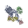
| Title | Crystal structure of the measles virus hemagglutinin bound to its cellular receptor SLAM (Form I, MV-H-SLAM(N102H/R108Y) fusion) | ||||||
 Components Components | Hemagglutinin, CDw150 | ||||||
 Keywords Keywords | viral protein/membrane protein / viral protein-receptor complex / six-bladed beta-propeller fold / Immunoglobulin fold / beta-sandwich / viral protein-membrane protein complex | ||||||
| Function / homology |  Function and homology information Function and homology informationlymphocyte activation / negative regulation of cytokine production / phagocytosis / signaling receptor activity / adaptive immune response / cell adhesion / host cell surface receptor binding / receptor ligand activity / innate immune response / viral envelope ...lymphocyte activation / negative regulation of cytokine production / phagocytosis / signaling receptor activity / adaptive immune response / cell adhesion / host cell surface receptor binding / receptor ligand activity / innate immune response / viral envelope / positive regulation of cell population proliferation / symbiont entry into host cell / virion attachment to host cell / host cell plasma membrane / virion membrane / cell surface / identical protein binding / plasma membrane Similarity search - Function | ||||||
| Biological species |   Measles virus Measles virussynthetic construct (others)  Saguinus oedipus (cotton-top tamarin) Saguinus oedipus (cotton-top tamarin) | ||||||
| Method |  X-RAY DIFFRACTION / SYNCHROTRON / MOLECULAR REPLACEMENT / Resolution: 3.55 Å X-RAY DIFFRACTION / SYNCHROTRON / MOLECULAR REPLACEMENT / Resolution: 3.55 Å | ||||||
 Authors Authors | Hashiguchi, T. / Ose, T. / Kubota, M. / Maita, N. / Kamishikiryo, J. / Maenaka, K. / Yanagi, Y. | ||||||
 Citation Citation | Journal: Nat.Struct.Mol.Biol. / Year: 2011 Title: Structure of the measles virus hemagglutinin bound to its cellular receptor SLAM Authors: Hashiguchi, T. / Ose, T. / Kubota, M. / Maita, N. / Kamishikiryo, J. / Maenaka, K. / Yanagi, Y. | ||||||
| History |
|
- Structure visualization
| Structure viewer | Molecule: MolmilJmol/JSmol |
|---|
- Downloads & links
-Download
| PDBx/mmCIF format | 3alw.cif.gz | 117.5 KB | Display | PDBx/mmCIF format |
|---|---|---|---|---|
| PDB format | pdb3alw.ent.gz | 88.9 KB | Display | PDB format |
| PDBx/mmJSON format | 3alw.json.gz | Tree view | PDBx/mmJSON format | |
| Others |  Other downloads Other downloads |
-Validation report
| Arichive directory | https://data.pdbj.org/pub/pdb/validation_reports/al/3alwftp://data.pdbj.org/pub/pdb/validation_reports/al/3alw | HTTPS FTP |
|---|
-Related structure data
| Related structure data | 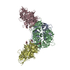 3alxC 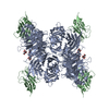 3alzC 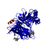 2zb6S C: citing same article ( S: Starting model for refinement |
|---|---|
| Similar structure data |


-Links
 PDBj
PDBj

- Assembly
| Deposited unit | 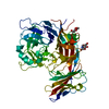
| ||||||||
|---|---|---|---|---|---|---|---|---|---|
| 1 | 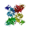
| ||||||||
| 2 | x 6
| ||||||||
| Unit cell |
|
-Components
| #1: Protein | Mass: 62078.816 Da / Num. of mol.: 1 Fragment: Hemagglutinin head domain/CD150 V domain, UNP reisudes 30-140 Mutation: N692H, R698Y (CDw150) Source method: isolated from a genetically manipulated source Details: MV-H/SLAM fusion protein Source: (gene. exp.) Measles virus, (gene. exp.) synthetic construct (others), (gene. exp.) Saguinus oedipus (cotton-top tamarin)Strain: Edmonston B, B95a / Gene: Hemagglutinin, CD150 / Plasmid: pCA7 / Cell line (production host): HEK293SGnTI(-) / Production host:  Homo sapiens (human) Homo sapiens (human)References: UniProt: E2RZS2, UniProt: Q9GJT3, UniProt: P08362*PLUS | ||||
|---|---|---|---|---|---|
| #2: Sugar |   Type: D-saccharide, beta linking / Mass: 221.208 Da / Num. of mol.: 2 Type: D-saccharide, beta linking / Mass: 221.208 Da / Num. of mol.: 2Source method: isolated from a genetically manipulated source Formula: C8H15NO6 Has protein modification | Y | Sequence details | THE GGGSGGGSGG | |
-Experimental details
-Experiment
| Experiment | Method: X-RAY DIFFRACTION / Number of used crystals: 1 |
|---|
- Sample preparation
| Crystal | Density Matthews: 9.990449 Å3/Da / Density % sol: 87.68824 % |
|---|---|
| Crystal grow | Temperature: 293 K / Method: vapor diffusion, floating drop / pH: 5.8 Details: 0.1M imidazole pH5.8, 0.5M (NH4)2SO4, 0.7M Li2SO4, 3% ethylene glycol, VAPOR DIFFUSION, FLOATING DROP, temperature 293K |
-Data collection
| Diffraction | Mean temperature: 100 K |
|---|---|
| Diffraction source | Source: SYNCHROTRON / Site: SPring-8  / Beamline: BL41XU / Wavelength: 1 Å / Beamline: BL41XU / Wavelength: 1 Å |
| Detector | Type: ADSC QUANTUM 315 / Detector: CCD / Date: Jul 22, 2009 |
| Radiation | Protocol: SINGLE WAVELENGTH / Monochromatic (M) / Laue (L): M / Scattering type: x-ray |
| Radiation wavelength | Wavelength: 1 Å / Relative weight: 1 |
| Reflection | Resolution: 3.55→30 Å / Num. obs: 28716 / % possible obs: 99.5 % / Observed criterion σ(F): 0 / Observed criterion σ(I): 0 / Redundancy: 20 % / Biso Wilson estimate: 78.3 Å2 / Rmerge(I) obs: 0.074 / Net I/σ(I): 38.6 |
| Reflection shell | Resolution: 3.55→3.68 Å / Redundancy: 20 % / Rmerge(I) obs: 0.498 / Mean I/σ(I) obs: 6.5 / Num. unique all: 2831 / % possible all: 100 |
- Processing
| Software |
| ||||||||||||||||||||||||||||||||||||||||||||||||||||||||||||
|---|---|---|---|---|---|---|---|---|---|---|---|---|---|---|---|---|---|---|---|---|---|---|---|---|---|---|---|---|---|---|---|---|---|---|---|---|---|---|---|---|---|---|---|---|---|---|---|---|---|---|---|---|---|---|---|---|---|---|---|---|---|
| Refinement | Method to determine structure: MOLECULAR REPLACEMENT Starting model: PDB ENTRY 2ZB6 Resolution: 3.55→29.89 Å / Rfactor Rfree error: 0.007 / Data cutoff high absF: 4911388.71 / Data cutoff low absF: 0 / Isotropic thermal model: GROUP / Cross valid method: THROUGHOUT / σ(F): 0 / Stereochemistry target values: Engh & Huber / Details: BULK SOLVENT MODEL USED
| ||||||||||||||||||||||||||||||||||||||||||||||||||||||||||||
| Solvent computation | Solvent model: FLAT MODEL / Bsol: 99.3884 Å2 / ksol: 0.3 e/Å3 | ||||||||||||||||||||||||||||||||||||||||||||||||||||||||||||
| Displacement parameters | Biso mean: 138.9 Å2
| ||||||||||||||||||||||||||||||||||||||||||||||||||||||||||||
| Refine analyze |
| ||||||||||||||||||||||||||||||||||||||||||||||||||||||||||||
| Refinement step | Cycle: LAST / Resolution: 3.55→29.89 Å
| ||||||||||||||||||||||||||||||||||||||||||||||||||||||||||||
| Refine LS restraints |
| ||||||||||||||||||||||||||||||||||||||||||||||||||||||||||||
| LS refinement shell | Resolution: 3.55→3.68 Å / Rfactor Rfree error: 0.029 / Total num. of bins used: 10
| ||||||||||||||||||||||||||||||||||||||||||||||||||||||||||||
| Xplor file |
|
