 Movie
Movie Controller


+ Open data

- Basic information
| Entry | Database: PDB / ID: 6lys | ||||||
|---|---|---|---|---|---|---|---|
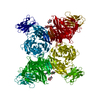
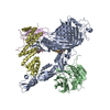
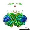
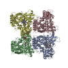
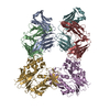
| Title | Structure of the BAM complex | ||||||
 Components Components |
| ||||||
 Keywords Keywords | MEMBRANE PROTEIN / b-barrel assembly machinery (BAM) complex | ||||||
| Function / homology |  Function and homology information Function and homology informationBam protein complex / Gram-negative-bacterium-type cell outer membrane assembly / protein insertion into membrane / Secretion of toxins / cell outer membrane / protein-folding chaperone binding / protein-macromolecule adaptor activity / cell adhesion / response to antibiotic / cell surface ...Bam protein complex / Gram-negative-bacterium-type cell outer membrane assembly / protein insertion into membrane / Secretion of toxins / cell outer membrane / protein-folding chaperone binding / protein-macromolecule adaptor activity / cell adhesion / response to antibiotic / cell surface / membrane / identical protein binding Similarity search - Function | ||||||
| Biological species |  | ||||||
| Method |  X-RAY DIFFRACTION / SYNCHROTRON / MOLECULAR REPLACEMENT / Resolution: 3.05 Å X-RAY DIFFRACTION / SYNCHROTRON / MOLECULAR REPLACEMENT / Resolution: 3.05 Å | ||||||
 Authors Authors | Xiao, L. / Huang, Y. | ||||||
 Citation Citation | Journal: FASEB J / Year: 2021 Title: Structures of the β-barrel assembly machine recognizing outer membrane protein substrates. Authors: Le Xiao / Long Han / Bufan Li / Manfeng Zhang / Haizhen Zhou / Qingshan Luo / Xinzheng Zhang / Yihua Huang /  Abstract: β-barrel outer membrane proteins (β-OMPs) play critical roles in nutrition acquisition, protein import/export, and other fundamental biological processes. The assembly of β-OMPs in Gram-negative ...β-barrel outer membrane proteins (β-OMPs) play critical roles in nutrition acquisition, protein import/export, and other fundamental biological processes. The assembly of β-OMPs in Gram-negative bacteria is mediated by the β-barrel assembly machinery (BAM) complex, yet its precise mechanism remains elusive. Here, we report two structures of the BAM complex in detergents and in nanodisks, and two crystal structures of the BAM complex with bound substrates. Structural analysis indicates that the membrane compositions surrounding the BAM complex could modulate its overall conformations, indicating low energy barriers between different conformational states and a highly dynamic nature of the BAM complex. Importantly, structures of the BAM complex with bound substrates and the related functional analysis show that the first β-strand of the BamA β-barrel (β1 ) in the BAM complex is associated with the last but not the first β-strand of a β-OMP substrate via antiparallel β-strand interactions. These observations are consistent with the β-signal hypothesis during β-OMP biogenesis, and suggest that the β1 strand in the BAM complex may interact with the last β-strand of an incoming β-OMP substrate upon their release from the chaperone-bound state. | ||||||
| History |
|
- Structure visualization
| Structure viewer | Molecule: MolmilJmol/JSmol |
|---|
- Downloads & links
-Download
| PDBx/mmCIF format | 6lys.cif.gz | 320.6 KB | Display | PDBx/mmCIF format |
|---|---|---|---|---|
| PDB format | pdb6lys.ent.gz | 251.2 KB | Display | PDB format |
| PDBx/mmJSON format | 6lys.json.gz | Tree view | PDBx/mmJSON format | |
| Others |  Other downloads Other downloads |
-Validation report
| Arichive directory | https://data.pdbj.org/pub/pdb/validation_reports/ly/6lysftp://data.pdbj.org/pub/pdb/validation_reports/ly/6lys | HTTPS FTP |
|---|
-Related structure data
| Related structure data |  6lyqC  6lyrC 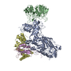 6lyuC 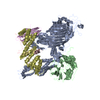 5d0oS S: Starting model for refinement C: citing same article ( |
|---|---|
| Similar structure data |


-Links
 PDBj
PDBj


- Assembly
| Deposited unit | 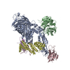
| ||||||||
|---|---|---|---|---|---|---|---|---|---|
| 1 |
| ||||||||
| Unit cell |
|
-Components
| #1: Protein | Mass: 90643.383 Da / Num. of mol.: 1 Source method: isolated from a genetically manipulated source Source: (gene. exp.) Gene: bamA, yaeT, yzzN, yzzY, b0177, JW0172 / Production host: |
|---|---|
| #2: Protein | Mass: 42961.086 Da / Num. of mol.: 1 Source method: isolated from a genetically manipulated source Source: (gene. exp.) Gene: bamB, yfgL, b2512, JW2496 / Production host: |
| #3: Protein | Mass: 27858.350 Da / Num. of mol.: 1 Source method: isolated from a genetically manipulated source Source: (gene. exp.) Gene: bamD, yfiO, b2595, JW2577 / Production host: |
| #4: Protein | Mass: 13139.857 Da / Num. of mol.: 1 Source method: isolated from a genetically manipulated source Source: (gene. exp.) Gene: bamE, smpA, b2617, JW2598 / Production host: |
| #5: Protein | Mass: 36875.277 Da / Num. of mol.: 1 Source method: isolated from a genetically manipulated source Source: (gene. exp.) Gene: bamC, dapX, nlpB, b2477, JW2462 / Production host: |
| Has protein modification | Y |
-Experimental details
-Experiment
| Experiment | Method: X-RAY DIFFRACTION / Number of used crystals: 1 |
|---|
- Sample preparation
| Crystal | Density Matthews: 3.48 Å3/Da / Density % sol: 64.62 % |
|---|---|
| Crystal grow | Temperature: 289 K / Method: vapor diffusion, hanging drop / Details: 27-30% PEG 400, 100mM NaCl |
-Data collection
| Diffraction | Mean temperature: 100 K / Serial crystal experiment: N |
|---|---|
| Diffraction source | Source: SYNCHROTRON / Site: SSRF / Beamline: BL17U / Wavelength: 0.97919 Å |
| Detector | Type: DECTRIS PILATUS3 S 6M / Detector: PIXEL / Date: Dec 15, 2018 |
| Radiation | Protocol: SINGLE WAVELENGTH / Monochromatic (M) / Laue (L): M / Scattering type: x-ray |
| Radiation wavelength | Wavelength: 0.97919 Å / Relative weight: 1 |
| Reflection | Resolution: 3.05→50 Å / Num. obs: 53217 / % possible obs: 99.9 % / Redundancy: 12.8 % / CC1/2: 0.998 / Net I/σ(I): 20.7 |
| Reflection shell | Resolution: 3.05→3.16 Å / Num. unique obs: 3492 / CC1/2: 0.904 |
- Processing
| Software |
| ||||||||||||||||||||
|---|---|---|---|---|---|---|---|---|---|---|---|---|---|---|---|---|---|---|---|---|---|
| Refinement | Method to determine structure: MOLECULAR REPLACEMENT Starting model: 5D0O Resolution: 3.05→10 Å / Cor.coef. Fo:Fc: 0.928 / Cor.coef. Fo:Fc free: 0.896 / SU B: 20.651 / SU ML: 0.352 / Cross valid method: THROUGHOUT / σ(F): 0 / ESU R: 1.266 / ESU R Free: 0.428 / Stereochemistry target values: MAXIMUM LIKELIHOOD Details: HYDROGENS HAVE BEEN ADDED IN THE RIDING POSITIONS U VALUES : REFINED INDIVIDUALLY
| ||||||||||||||||||||
| Solvent computation | Ion probe radii: 0.8 Å / Shrinkage radii: 0.8 Å / VDW probe radii: 1.2 Å / Solvent model: MASK | ||||||||||||||||||||
| Displacement parameters | Biso max: 209.75 Å2 / Biso mean: 91.413 Å2 / Biso min: 62.66 Å2
| ||||||||||||||||||||
| Refinement step | Cycle: final / Resolution: 3.05→10 Å
| ||||||||||||||||||||
| LS refinement shell | Resolution: 3.051→3.123 Å / Rfactor Rfree error: 0 / Total num. of bins used: 20
|
