 Movie
Movie Controller

[English] 日本語
 Yorodumi
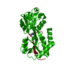
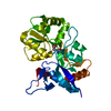
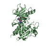
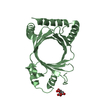
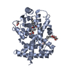
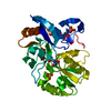
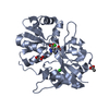
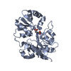
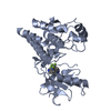
- PDB-1txf: CRYSTAL STRUCTURE OF THE GLUR5 LIGAND BINDING CORE IN COMPLEX WIT... -
+ Open data

- Basic information
| Entry | Database: PDB / ID: 1txf | ||||||
|---|---|---|---|---|---|---|---|
| Title | CRYSTAL STRUCTURE OF THE GLUR5 LIGAND BINDING CORE IN COMPLEX WITH GLUTAMATE AT 2.1 ANGSTROM RESOLUTION | ||||||
 Components Components | Glutamate receptor, ionotropic kainate 1 | ||||||
 Keywords Keywords | MEMBRANE PROTEIN | ||||||
| Function / homology |  Function and homology information Function and homology informationnegative regulation of synaptic transmission, GABAergic / L-glutamate transmembrane transporter activity / positive regulation of gamma-aminobutyric acid secretion / Activation of Na-permeable kainate receptors / Activation of Ca-permeable Kainate Receptor / kainate selective glutamate receptor complex / regulation of short-term neuronal synaptic plasticity / negative regulation of synaptic transmission, glutamatergic / glutamate binding / inhibitory postsynaptic potential ...negative regulation of synaptic transmission, GABAergic / L-glutamate transmembrane transporter activity / positive regulation of gamma-aminobutyric acid secretion / Activation of Na-permeable kainate receptors / Activation of Ca-permeable Kainate Receptor / kainate selective glutamate receptor complex / regulation of short-term neuronal synaptic plasticity / negative regulation of synaptic transmission, glutamatergic / glutamate binding / inhibitory postsynaptic potential / adult behavior / kainate selective glutamate receptor activity / behavioral response to pain / extracellularly glutamate-gated ion channel activity / modulation of excitatory postsynaptic potential / ionotropic glutamate receptor complex / membrane depolarization / glutamate-gated receptor activity / establishment of localization in cell / glutamate-gated calcium ion channel activity / ionotropic glutamate receptor signaling pathway / ligand-gated monoatomic ion channel activity involved in regulation of presynaptic membrane potential / presynaptic modulation of chemical synaptic transmission / positive regulation of synaptic transmission, GABAergic / SNARE binding / synaptic transmission, glutamatergic / transmitter-gated monoatomic ion channel activity involved in regulation of postsynaptic membrane potential / regulation of membrane potential / excitatory postsynaptic potential / regulation of synaptic plasticity / postsynaptic density membrane / modulation of chemical synaptic transmission / terminal bouton / nervous system development / presynaptic membrane / scaffold protein binding / chemical synaptic transmission / postsynaptic membrane / signaling receptor complex / postsynaptic density / neuronal cell body / synapse / dendrite / glutamatergic synapse / membrane / identical protein binding / plasma membrane Similarity search - Function | ||||||
| Biological species |  | ||||||
| Method |  X-RAY DIFFRACTION / SYNCHROTRON / MOLECULAR REPLACEMENT / Resolution: 2.1 Å X-RAY DIFFRACTION / SYNCHROTRON / MOLECULAR REPLACEMENT / Resolution: 2.1 Å | ||||||
 Authors Authors | Mayer, M.L. | ||||||
 Citation Citation | Journal: Neuron / Year: 2005 Title: Crystal structures of the GluR5 and GluR6 ligand binding cores: Molecular mechanisms underlying kainate receptor selectivity Authors: Mayer, M.L. | ||||||
| History |
| ||||||
| Remark 999 | SEQUENCE THE NATIVE GLUR-5 IS A MEMBRANE PROTEIN. THE PROTEIN CRYSTALLIZED BY THE AUTHOR IS THE ...SEQUENCE THE NATIVE GLUR-5 IS A MEMBRANE PROTEIN. THE PROTEIN CRYSTALLIZED BY THE AUTHOR IS THE EXTRACELLULAR LIGAND BINDING DOMAIN OF GLUR-5. TRANSMEMBRANE REGIONS WERE GENETICALLY REMOVED AND REPLACED WITH A GLY-THR LINKER. THE SEQUENCE, AS A RESULT, MATCHES DISCONTINUOUSLY WITH THE REFERENCE DATABASE | ||||||
| Remark 300 | BIOMOLECULE THE BIOLOGICAL UNIT IS BELIEVED TO BE A HETEROTETRAMER, THERE IS ONLY ONE MOLECULE IN ...BIOMOLECULE THE BIOLOGICAL UNIT IS BELIEVED TO BE A HETEROTETRAMER, THERE IS ONLY ONE MOLECULE IN THE ASYMMETRIC UNIT AND SYMMETRY OPERATIONS CANNOT BE APPLIED TO GENERATE THE TETRAMER. |
- Structure visualization
| Structure viewer | Molecule: MolmilJmol/JSmol |
|---|
- Downloads & links
-Download
| PDBx/mmCIF format | 1txf.cif.gz | 64.8 KB | Display | PDBx/mmCIF format |
|---|---|---|---|---|
| PDB format | pdb1txf.ent.gz | 46.7 KB | Display | PDB format |
| PDBx/mmJSON format | 1txf.json.gz | Tree view | PDBx/mmJSON format | |
| Others |  Other downloads Other downloads |
-Validation report
| Arichive directory | https://data.pdbj.org/pub/pdb/validation_reports/tx/1txfftp://data.pdbj.org/pub/pdb/validation_reports/tx/1txf | HTTPS FTP |
|---|
-Related structure data
| Related structure data |  1s50C  1s7ySC  1s9tC  1sd3C  1tt1C S: Starting model for refinement C: citing same article ( |
|---|---|
| Similar structure data |


-Links
 PDBj
PDBj


- Assembly
| Deposited unit | 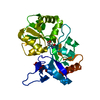
| ||||||||
|---|---|---|---|---|---|---|---|---|---|
| 1 |
| ||||||||
| Unit cell |
|
-Components
| #1: Protein | Mass: 29253.566 Da / Num. of mol.: 1 Fragment: GluR5 ligand binding core (sequence database 446-559 and 682-821) Source method: isolated from a genetically manipulated source Source: (gene. exp.)  |
|---|---|
| #2: Chemical | ChemComp-GLU /   Type: L-peptide linking / Mass: 147.129 Da / Num. of mol.: 1 / Source method: obtained synthetically / Formula: C5H9NO4 Type: L-peptide linking / Mass: 147.129 Da / Num. of mol.: 1 / Source method: obtained synthetically / Formula: C5H9NO4 |
| #3: Water | ChemComp-HOH /  Mass: 18.015 Da / Num. of mol.: 93 / Source method: isolated from a natural source / Formula: H2O Mass: 18.015 Da / Num. of mol.: 93 / Source method: isolated from a natural source / Formula: H2O |
-Experimental details
-Experiment
| Experiment | Method: X-RAY DIFFRACTION / Number of used crystals: 1 |
|---|
- Sample preparation
| Crystal | Density Matthews: 3.1 Å3/Da / Density % sol: 60.4 % |
|---|---|
| Crystal grow | Temperature: 293 K / Method: vapor diffusion, hanging drop / pH: 7.5 Details: 20% PEG 10K, 100 mM HEPES, 20 mM NaCl, 1 mM EDTA, 10 mM Glutamate, pH 7.5, VAPOR DIFFUSION, HANGING DROP, temperature 293K |
-Data collection
| Diffraction | Mean temperature: 100 K |
|---|---|
| Diffraction source | Source: SYNCHROTRON / Site: APS  / Beamline: 22-ID / Wavelength: 0.99997 Å / Beamline: 22-ID / Wavelength: 0.99997 Å |
| Detector | Type: MARRESEARCH / Detector: CCD / Date: Nov 1, 2003 |
| Radiation | Protocol: SINGLE WAVELENGTH / Monochromatic (M) / Laue (L): M / Scattering type: x-ray |
| Radiation wavelength | Wavelength: 0.99997 Å / Relative weight: 1 |
| Reflection | Resolution: 2.1→50 Å / Num. all: 21345 / Num. obs: 21345 / % possible obs: 84.3 % / Observed criterion σ(F): 1 / Observed criterion σ(I): 2 / Redundancy: 2.5 % / Biso Wilson estimate: 39.9 Å2 / Rmerge(I) obs: 0.045 / Net I/σ(I): 12.7 |
| Reflection shell | Resolution: 2.1→2.18 Å / Redundancy: 2.2 % / Rmerge(I) obs: 0.269 / Mean I/σ(I) obs: 2.2 / Num. unique all: 1042 / % possible all: 49.7 |
- Processing
| Software |
| |||||||||||||||||||||||||
|---|---|---|---|---|---|---|---|---|---|---|---|---|---|---|---|---|---|---|---|---|---|---|---|---|---|---|
| Refinement | Method to determine structure: MOLECULAR REPLACEMENT Starting model: PDB ENTRY 1S7Y Resolution: 2.1→32.4 Å / Rfactor Rfree error: 0.007 / Data cutoff high absF: 1282416.77 / Data cutoff low absF: 0 / Isotropic thermal model: RESTRAINED / Cross valid method: THROUGHOUT / σ(F): 0 / Stereochemistry target values: Engh & Huber
| |||||||||||||||||||||||||
| Solvent computation | Solvent model: FLAT MODEL / Bsol: 44.2134 Å2 / ksol: 0.3641 e/Å3 | |||||||||||||||||||||||||
| Displacement parameters | Biso mean: 41.8 Å2
| |||||||||||||||||||||||||
| Refine analyze |
| |||||||||||||||||||||||||
| Refinement step | Cycle: LAST / Resolution: 2.1→32.4 Å
| |||||||||||||||||||||||||
| Refine LS restraints |
| |||||||||||||||||||||||||
| LS refinement shell | Resolution: 2.1→2.23 Å / Rfactor Rfree error: 0.028 / Total num. of bins used: 6
| |||||||||||||||||||||||||
| Xplor file |
|
