 Movie
Movie Controller

[English] 日本語
 Yorodumi
- PDB-1ii5: CRYSTAL STRUCTURE OF THE GLUR0 LIGAND BINDING CORE COMPLEX WITH L... -
+ Open data

- Basic information
| Entry | Database: PDB / ID: 1ii5 | ||||||
|---|---|---|---|---|---|---|---|
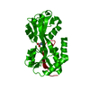
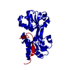
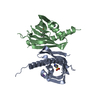
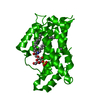
| Title | CRYSTAL STRUCTURE OF THE GLUR0 LIGAND BINDING CORE COMPLEX WITH L-GLUTAMATE | ||||||
 Components Components | Slr1257 protein | ||||||
 Keywords Keywords | MEMBRANE PROTEIN | ||||||
| Function / homology |  Function and homology information Function and homology information | ||||||
| Biological species |  | ||||||
| Method |  X-RAY DIFFRACTION / SYNCHROTRON / MAD / Resolution: 1.6 Å X-RAY DIFFRACTION / SYNCHROTRON / MAD / Resolution: 1.6 Å | ||||||
 Authors Authors | Mayer, M.L. / Olson, R. / Gouaux, E. | ||||||
 Citation Citation | Journal: J.Mol.Biol. / Year: 2001 Title: Mechanisms for ligand binding to GluR0 ion channels: crystal structures of the glutamate and serine complexes and a closed apo state. Authors: Mayer, M.L. / Olson, R. / Gouaux, E. #1: Journal: Nature / Year: 1999Title: FUNCTIONAL CHARACTERIZATION OF A POTASSIUM-SELECTIVE PROKARYOTIC GLUTAMATE RECEPTOR Authors: CHEN, G.-Q. / CUI, C. / MAYER, M.L. / GOUAUX, E. | ||||||
| History |
| ||||||
| Remark 999 | SEQUENCE NATIVE GLURO IS A MEMBRANE PROTEIN. THE PROTEIN CRYSTALLIZED BY THE AUTHOR IS THE ...SEQUENCE NATIVE GLURO IS A MEMBRANE PROTEIN. THE PROTEIN CRYSTALLIZED BY THE AUTHOR IS THE EXTRACELLULAR LIGAND BINDING DOMAIN OF GLURO. TRANSMEMBRANE REGIONS WERE GENETICALLY REMOVED AND REPLACED WITH A THR LINKER. THE SEQUENCE, AS A RESULT, MATCHES DISCONTINUOUSLY WITH THE REFERENCE DATABASE. |
- Structure visualization
| Structure viewer | Molecule: MolmilJmol/JSmol |
|---|
- Downloads & links
-Download
| PDBx/mmCIF format | 1ii5.cif.gz | 57.4 KB | Display | PDBx/mmCIF format |
|---|---|---|---|---|
| PDB format | pdb1ii5.ent.gz | 40.8 KB | Display | PDB format |
| PDBx/mmJSON format | 1ii5.json.gz | Tree view | PDBx/mmJSON format | |
| Others |  Other downloads Other downloads |
-Validation report
| Arichive directory | https://data.pdbj.org/pub/pdb/validation_reports/ii/1ii5ftp://data.pdbj.org/pub/pdb/validation_reports/ii/1ii5 | HTTPS FTP |
|---|
-Related structure data


-Links
 PDBj
PDBj


- Assembly
| Deposited unit | 
| ||||||||
|---|---|---|---|---|---|---|---|---|---|
| 1 | 
| ||||||||
| Unit cell |
|
-Components
| #1: Protein | Mass: 25694.107 Da / Num. of mol.: 1 Fragment: GluR0 ligand binding core, residues 44-140, 256-385 Source method: isolated from a genetically manipulated source Source: (gene. exp.) Strain: PCC 6803 / Kazusa / Gene: GluR0 slr1257, slr1257 / Species (production host): Escherichia coli / Production host: |
|---|---|
| #2: Chemical | ChemComp-GLU /   Type: L-peptide linking / Mass: 147.129 Da / Num. of mol.: 1 / Source method: obtained synthetically / Formula: C5H9NO4 Type: L-peptide linking / Mass: 147.129 Da / Num. of mol.: 1 / Source method: obtained synthetically / Formula: C5H9NO4 |
| #3: Water | ChemComp-HOH /  Mass: 18.015 Da / Num. of mol.: 134 / Source method: isolated from a natural source / Formula: H2O Mass: 18.015 Da / Num. of mol.: 134 / Source method: isolated from a natural source / Formula: H2O |
-Experimental details
-Experiment
| Experiment | Method: X-RAY DIFFRACTION / Number of used crystals: 1 |
|---|
- Sample preparation
| Crystal | Density Matthews: 2.35 Å3/Da / Density % sol: 47.57 % | |||||||||||||||||||||||||
|---|---|---|---|---|---|---|---|---|---|---|---|---|---|---|---|---|---|---|---|---|---|---|---|---|---|---|
| Crystal grow | Temperature: 277 K / Method: vapor diffusion, hanging drop / pH: 4.7 Details: 38% MPD, 100 mM sodium acetate, 10 mM L-glutamic acid, pH 4.7, VAPOR DIFFUSION, HANGING DROP, temperature 277K | |||||||||||||||||||||||||
| Crystal grow | *PLUS Temperature: 4 ℃ / PH range low: 4.8 / PH range high: 4.7 | |||||||||||||||||||||||||
| Components of the solutions | *PLUS
|
-Data collection
| Diffraction | Mean temperature: 110 K |
|---|---|
| Diffraction source | Source: SYNCHROTRON / Site: NSLS  / Beamline: X4A / Wavelength: 0.97625 Å / Beamline: X4A / Wavelength: 0.97625 Å |
| Detector | Type: ADSC QUANTUM 4 / Detector: CCD / Date: Jun 16, 2000 |
| Radiation | Protocol: SINGLE WAVELENGTH / Monochromatic (M) / Laue (L): M / Scattering type: x-ray |
| Radiation wavelength | Wavelength: 0.97625 Å / Relative weight: 1 |
| Reflection | Resolution: 1.6→30 Å / Num. obs: 31427 / % possible obs: 99.9 % / Observed criterion σ(F): 2 / Observed criterion σ(I): 2 / Redundancy: 5.7 % / Biso Wilson estimate: 20.4 Å2 / Rmerge(I) obs: 0.055 / Net I/σ(I): 22.8 |
| Reflection shell | Resolution: 1.6→1.66 Å / Redundancy: 2.8 % / Rmerge(I) obs: 0.12 / Mean I/σ(I) obs: 8.4 / Num. unique all: 3130 / % possible all: 99.8 |
| Reflection | *PLUS Lowest resolution: 20 Å / Num. measured all: 179108 |
| Reflection shell | *PLUS % possible obs: 99.8 % / Rmerge(I) obs: 0.12 |
- Processing
| Software |
| ||||||||||||||||||||
|---|---|---|---|---|---|---|---|---|---|---|---|---|---|---|---|---|---|---|---|---|---|
| Refinement | Method to determine structure: MAD / Resolution: 1.6→20 Å / Cross valid method: THROUGHOUT / σ(F): 2 / Stereochemistry target values: Engh & Huber
| ||||||||||||||||||||
| Refinement step | Cycle: LAST / Resolution: 1.6→20 Å
| ||||||||||||||||||||
| Refine LS restraints |
| ||||||||||||||||||||
| LS refinement shell | Resolution: 1.6→1.67 Å
| ||||||||||||||||||||
| Software | *PLUS Name: X-PLOR / Version: 3.851 / Classification: refinement | ||||||||||||||||||||
| Refinement | *PLUS Lowest resolution: 30 Å / σ(F): 2 | ||||||||||||||||||||
| Solvent computation | *PLUS | ||||||||||||||||||||
| Displacement parameters | *PLUS | ||||||||||||||||||||
| Refine LS restraints | *PLUS
| ||||||||||||||||||||
| LS refinement shell | *PLUS Rfactor Rfree: 0.377 / Rfactor Rwork: 0.334 |
