 Movie
Movie Controller

[English] 日本語
 Yorodumi
- PDB-4up2: Crystal structure of Escherichia coli tryptophanase purified from... -
+ Open data

- Basic information
| Entry | Database: PDB / ID: 4up2 | |||||||||
|---|---|---|---|---|---|---|---|---|---|---|
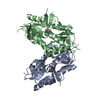
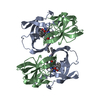
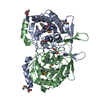
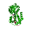
| Title | Crystal structure of Escherichia coli tryptophanase purified from alkaline stressed bacterial culture. | |||||||||
 Components Components | TRYPTOPHANASE | |||||||||
 Keywords Keywords | LYASE / ALKALINE STRESS / PROTEIN PURIFICATION | |||||||||
| Function / homology |  Function and homology information Function and homology information | |||||||||
| Biological species |  | |||||||||
| Method |  X-RAY DIFFRACTION / SYNCHROTRON / MOLECULAR REPLACEMENT / Resolution: 2.78 Å X-RAY DIFFRACTION / SYNCHROTRON / MOLECULAR REPLACEMENT / Resolution: 2.78 Å | |||||||||
 Authors Authors | Rety, S. / Deschamps, P. / Leulliot, N. | |||||||||
 Citation Citation | Journal: Acta Crystallogr.,Sect.F / Year: 2015 Title: Structure of Escherichia Coli Tryptophanase Purified from an Alkaline-Stressed Bacterial Culture. Authors: Rety, S. / Deschamps, P. / Leulliot, N. | |||||||||
| History |
|
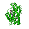
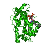
- Structure visualization
| Structure viewer | Molecule: MolmilJmol/JSmol |
|---|
- Downloads & links
-Download
| PDBx/mmCIF format | 4up2.cif.gz | 598.4 KB | Display | PDBx/mmCIF format |
|---|---|---|---|---|
| PDB format | pdb4up2.ent.gz | 505.6 KB | Display | PDB format |
| PDBx/mmJSON format | 4up2.json.gz | Tree view | PDBx/mmJSON format | |
| Others |  Other downloads Other downloads |
-Validation report
| Arichive directory | https://data.pdbj.org/pub/pdb/validation_reports/up/4up2ftp://data.pdbj.org/pub/pdb/validation_reports/up/4up2 | HTTPS FTP |
|---|
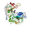
-Related structure data
| Related structure data | 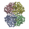 2c44S S: Starting model for refinement |
|---|---|
| Similar structure data |


-Links
 PDBj
PDBj


- Assembly
| Deposited unit | 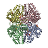
| ||||||||
|---|---|---|---|---|---|---|---|---|---|
| 1 | 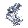
| ||||||||
| 2 | 
| ||||||||
| 3 | 
| ||||||||
| 4 | 
| ||||||||
| Unit cell |
|
-Components
| #1: Protein | Mass: 52850.266 Da / Num. of mol.: 4 / Fragment: RESIDUES 6-476 Source method: isolated from a genetically manipulated source Source: (gene. exp.) #2: Chemical | ChemComp-SO4 /   Mass: 96.063 Da / Num. of mol.: 28 / Source method: obtained synthetically / Formula: SO4 Mass: 96.063 Da / Num. of mol.: 28 / Source method: obtained synthetically / Formula: SO4#3: Chemical | ChemComp-BME / |   Mass: 78.133 Da / Num. of mol.: 1 / Source method: obtained synthetically / Formula: C2H6OS Mass: 78.133 Da / Num. of mol.: 1 / Source method: obtained synthetically / Formula: C2H6OS#4: Water | ChemComp-HOH / |  Mass: 18.015 Da / Num. of mol.: 113 / Source method: isolated from a natural source / Formula: H2O Mass: 18.015 Da / Num. of mol.: 113 / Source method: isolated from a natural source / Formula: H2O |
|---|
-Experimental details
-Experiment
| Experiment | Method: X-RAY DIFFRACTION / Number of used crystals: 1 |
|---|
- Sample preparation
| Crystal | Density Matthews: 3.32 Å3/Da / Density % sol: 63.02 % / Description: NONE |
|---|---|
| Crystal grow | Details: 25% PEG 4000 30MM AMMONIUM SULFATE 20MM B-MERCAPTOETHANOL 100MM TRIS-HCL PH 9 |
-Data collection
| Diffraction | Mean temperature: 100 K |
|---|---|
| Diffraction source | Source: SYNCHROTRON / Site: ESRF  / Beamline: ID23-1 / Wavelength: 0.97625 / Beamline: ID23-1 / Wavelength: 0.97625 |
| Detector | Type: ADSC QUANTUM 315r / Detector: CCD / Date: Sep 25, 2009 |
| Radiation | Protocol: SINGLE WAVELENGTH / Monochromatic (M) / Laue (L): M / Scattering type: x-ray |
| Radiation wavelength | Wavelength: 0.97625 Å / Relative weight: 1 |
| Reflection | Resolution: 2.78→47.01 Å / Num. obs: 72775 / % possible obs: 99.9 % / Observed criterion σ(I): 2 / Redundancy: 42.6 % / Biso Wilson estimate: 56.34 Å2 / Rmerge(I) obs: 0.175 / Net I/σ(I): 25.72 |
| Reflection shell | Resolution: 2.78→2.88 Å / Redundancy: 43.7 % / Rmerge(I) obs: 0.21 / Mean I/σ(I) obs: 2.54 / % possible all: 100 |
- Processing
| Software |
| |||||||||||||||||||||||||||||||||||||||||||||||||||||||||||||||||||||||||||||||||||||||||||||||||||||||||||||||||||||||||||||||||||||||||||||||||||||||||||||||||||||||||||||||||||||||||||||
|---|---|---|---|---|---|---|---|---|---|---|---|---|---|---|---|---|---|---|---|---|---|---|---|---|---|---|---|---|---|---|---|---|---|---|---|---|---|---|---|---|---|---|---|---|---|---|---|---|---|---|---|---|---|---|---|---|---|---|---|---|---|---|---|---|---|---|---|---|---|---|---|---|---|---|---|---|---|---|---|---|---|---|---|---|---|---|---|---|---|---|---|---|---|---|---|---|---|---|---|---|---|---|---|---|---|---|---|---|---|---|---|---|---|---|---|---|---|---|---|---|---|---|---|---|---|---|---|---|---|---|---|---|---|---|---|---|---|---|---|---|---|---|---|---|---|---|---|---|---|---|---|---|---|---|---|---|---|---|---|---|---|---|---|---|---|---|---|---|---|---|---|---|---|---|---|---|---|---|---|---|---|---|---|---|---|---|---|---|---|---|
| Refinement | Method to determine structure: MOLECULAR REPLACEMENT Starting model: PDB ENTRY 2C44 Resolution: 2.78→47.014 Å / SU ML: 0.37 / σ(F): 1.35 / Phase error: 23.83 / Stereochemistry target values: ML
| |||||||||||||||||||||||||||||||||||||||||||||||||||||||||||||||||||||||||||||||||||||||||||||||||||||||||||||||||||||||||||||||||||||||||||||||||||||||||||||||||||||||||||||||||||||||||||||
| Solvent computation | Shrinkage radii: 0.9 Å / VDW probe radii: 1.11 Å / Solvent model: FLAT BULK SOLVENT MODEL | |||||||||||||||||||||||||||||||||||||||||||||||||||||||||||||||||||||||||||||||||||||||||||||||||||||||||||||||||||||||||||||||||||||||||||||||||||||||||||||||||||||||||||||||||||||||||||||
| Displacement parameters | Biso mean: 60.9 Å2 | |||||||||||||||||||||||||||||||||||||||||||||||||||||||||||||||||||||||||||||||||||||||||||||||||||||||||||||||||||||||||||||||||||||||||||||||||||||||||||||||||||||||||||||||||||||||||||||
| Refinement step | Cycle: LAST / Resolution: 2.78→47.014 Å
| |||||||||||||||||||||||||||||||||||||||||||||||||||||||||||||||||||||||||||||||||||||||||||||||||||||||||||||||||||||||||||||||||||||||||||||||||||||||||||||||||||||||||||||||||||||||||||||
| Refine LS restraints |
| |||||||||||||||||||||||||||||||||||||||||||||||||||||||||||||||||||||||||||||||||||||||||||||||||||||||||||||||||||||||||||||||||||||||||||||||||||||||||||||||||||||||||||||||||||||||||||||
| LS refinement shell |
|
