 Movie
Movie Controller

[English] 日本語
 Yorodumi
- PDB-4gxd: Crystal structure of D48V mutant of human GLTP bound with 12:0 di... -
+ Open data

- Basic information
| Entry | Database: PDB / ID: 4gxd | ||||||
|---|---|---|---|---|---|---|---|
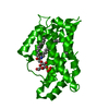
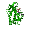
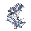
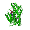
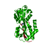
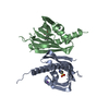
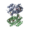
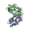
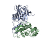
| Title | Crystal structure of D48V mutant of human GLTP bound with 12:0 disulfatide | ||||||
 Components Components | Glycolipid transfer protein | ||||||
 Keywords Keywords | LIPID TRANSPORT / GLTP-fold | ||||||
| Function / homology |  Function and homology information Function and homology informationlipid sensor activity / regulation of glucosylceramide biosynthetic process / glycolipid transfer activity / ceramide transport / ceramide 1-phosphate transfer activity / ceramide 1-phosphate binding / glycolipid binding / lipid transfer activity / Glycosphingolipid transport / intermembrane lipid transfer ...lipid sensor activity / regulation of glucosylceramide biosynthetic process / glycolipid transfer activity / ceramide transport / ceramide 1-phosphate transfer activity / ceramide 1-phosphate binding / glycolipid binding / lipid transfer activity / Glycosphingolipid transport / intermembrane lipid transfer / response to immobilization stress / lipid binding / identical protein binding / cytosol Similarity search - Function | ||||||
| Biological species |  Homo sapiens (human) Homo sapiens (human) | ||||||
| Method |  X-RAY DIFFRACTION / SYNCHROTRON / MOLECULAR REPLACEMENT / Resolution: 2.1 Å X-RAY DIFFRACTION / SYNCHROTRON / MOLECULAR REPLACEMENT / Resolution: 2.1 Å | ||||||
 Authors Authors | Samygina, V.R. / Ochoa-Lizarralde, B. / Popov, A.N. / Malinina, L. | ||||||
 Citation Citation | Journal: Acta Crystallogr.,Sect.D / Year: 2013 Title: Structural insights into lipid-dependent reversible dimerization of human GLTP. Authors: Samygina, V.R. / Ochoa-Lizarralde, B. / Popov, A.N. / Cabo-Bilbao, A. / Goni-de-Cerio, F. / Molotkovsky, J.G. / Patel, D.J. / Brown, R.E. / Malinina, L. | ||||||
| History |
|
- Structure visualization
| Structure viewer | Molecule: MolmilJmol/JSmol |
|---|
- Downloads & links
-Download
| PDBx/mmCIF format | 4gxd.cif.gz | 96.1 KB | Display | PDBx/mmCIF format |
|---|---|---|---|---|
| PDB format | pdb4gxd.ent.gz | 73.4 KB | Display | PDB format |
| PDBx/mmJSON format | 4gxd.json.gz | Tree view | PDBx/mmJSON format | |
| Others |  Other downloads Other downloads |
-Validation report
| Arichive directory | https://data.pdbj.org/pub/pdb/validation_reports/gx/4gxdftp://data.pdbj.org/pub/pdb/validation_reports/gx/4gxd | HTTPS FTP |
|---|
-Related structure data
| Related structure data |  4gh0C  4ghpC  4ghsC  4gixC  4gjqC  4gvtC  4gxgC  4h2zC C: citing same article ( |
|---|---|
| Similar structure data |

-Links
 PDBj
PDBj- Assembly
| Deposited unit | 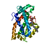
| ||||||||
|---|---|---|---|---|---|---|---|---|---|
| 1 | 
| ||||||||
| Unit cell |
|
-Components
| #1: Protein | Mass: 23861.820 Da / Num. of mol.: 1 / Mutation: D48V Source method: isolated from a genetically manipulated source Source: (gene. exp.) Homo sapiens (human) / Gene: GLTP / Production host:  |
|---|---|
| #2: Chemical | ChemComp-0SG /   Mass: 804.061 Da / Num. of mol.: 1 / Source method: obtained synthetically / Formula: C36H69NO14S2 Mass: 804.061 Da / Num. of mol.: 1 / Source method: obtained synthetically / Formula: C36H69NO14S2 |
| #3: Water | ChemComp-HOH /  Mass: 18.015 Da / Num. of mol.: 41 / Source method: isolated from a natural source / Formula: H2O Mass: 18.015 Da / Num. of mol.: 41 / Source method: isolated from a natural source / Formula: H2O |
-Experimental details
-Experiment
| Experiment | Method: X-RAY DIFFRACTION / Number of used crystals: 1 |
|---|
- Sample preparation
| Crystal | Density Matthews: 2 Å3/Da / Density % sol: 38.6 % |
|---|---|
| Crystal grow | Temperature: 298 K / Method: vapor diffusion, hanging drop / pH: 7 Details: 15% PEG 3350, 0.1M MES pH 7.0, VAPOR DIFFUSION, HANGING DROP, temperature 298K |
-Data collection
| Diffraction | Mean temperature: 100 K |
|---|---|
| Diffraction source | Source: SYNCHROTRON / Site: ESRF  / Beamline: ID2 / Wavelength: 0.9754 Å / Beamline: ID2 / Wavelength: 0.9754 Å |
| Detector | Type: ADSC QUANTUM 315 / Detector: CCD / Date: Sep 1, 2010 |
| Radiation | Monochromator: Si(111) / Protocol: SINGLE WAVELENGTH / Monochromatic (M) / Laue (L): M / Scattering type: x-ray |
| Radiation wavelength | Wavelength: 0.9754 Å / Relative weight: 1 |
| Reflection | Resolution: 2.1→20 Å / Num. all: 12893 / Num. obs: 12893 / % possible obs: 99.95 % / Observed criterion σ(F): 2 / Observed criterion σ(I): 2 |
| Reflection shell | Resolution: 2.1→2.15 Å / % possible all: 100 |
- Processing
| Software |
| ||||||||||||||||||||||||||||||||||||||||||||||||||||||||||||
|---|---|---|---|---|---|---|---|---|---|---|---|---|---|---|---|---|---|---|---|---|---|---|---|---|---|---|---|---|---|---|---|---|---|---|---|---|---|---|---|---|---|---|---|---|---|---|---|---|---|---|---|---|---|---|---|---|---|---|---|---|---|
| Refinement | Method to determine structure: MOLECULAR REPLACEMENT / Resolution: 2.1→14.76 Å / Cor.coef. Fo:Fc: 0.958 / Cor.coef. Fo:Fc free: 0.919 / SU B: 11.506 / SU ML: 0.156 / Cross valid method: THROUGHOUT / σ(F): 1 / ESU R: 0.264 / ESU R Free: 0.214 / Stereochemistry target values: MAXIMUM LIKELIHOOD / Details: HYDROGENS HAVE BEEN ADDED IN THE RIDING POSITIONS
| ||||||||||||||||||||||||||||||||||||||||||||||||||||||||||||
| Solvent computation | Ion probe radii: 0.8 Å / Shrinkage radii: 0.8 Å / VDW probe radii: 1.2 Å / Solvent model: MASK | ||||||||||||||||||||||||||||||||||||||||||||||||||||||||||||
| Displacement parameters | Biso mean: 44.068 Å2
| ||||||||||||||||||||||||||||||||||||||||||||||||||||||||||||
| Refinement step | Cycle: LAST / Resolution: 2.1→14.76 Å
| ||||||||||||||||||||||||||||||||||||||||||||||||||||||||||||
| Refine LS restraints |
| ||||||||||||||||||||||||||||||||||||||||||||||||||||||||||||
| LS refinement shell | Resolution: 2.1→2.154 Å / Total num. of bins used: 20
| ||||||||||||||||||||||||||||||||||||||||||||||||||||||||||||
| Refinement TLS params. | Method: refined / Origin x: -12.0207 Å / Origin y: -0.551 Å / Origin z: -13.4748 Å
|
