 Movie
Movie Controller

[English] 日本語
 Yorodumi
- PDB-2h5y: Crystallographic structure of the Molybdate-Binding Protein of Xa... -
+ Open data

- Basic information
| Entry | Database: PDB / ID: 2h5y | ||||||
|---|---|---|---|---|---|---|---|
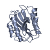
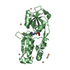
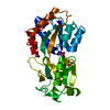
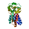
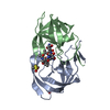
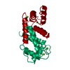
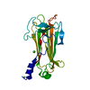
| Title | Crystallographic structure of the Molybdate-Binding Protein of Xanthomonas citri at 1.7 Ang resolution bound to molybdate | ||||||
 Components Components | Molybdate-binding periplasmic protein | ||||||
 Keywords Keywords | METAL TRANSPORT / Molybdate-binding protein / ModA / Xanthomonas axonopodis pv. citri | ||||||
| Function / homology |  Function and homology information Function and homology informationtungstate binding / molybdate ion binding / molybdate ion transport / outer membrane-bounded periplasmic space / metal ion binding Similarity search - Function | ||||||
| Biological species |  Xanthomonas axonopodis pv. citri (bacteria) Xanthomonas axonopodis pv. citri (bacteria) | ||||||
| Method |  X-RAY DIFFRACTION / SYNCHROTRON / MOLECULAR REPLACEMENT / Resolution: 1.7 Å X-RAY DIFFRACTION / SYNCHROTRON / MOLECULAR REPLACEMENT / Resolution: 1.7 Å | ||||||
 Authors Authors | Balan, A. / Santacruz, C.P. / Ferreira, L.C.S. / Barbosa, J.A.R.G. | ||||||
 Citation Citation | Journal: Acta Crystallogr.,Sect.F / Year: 2006 Title: Crystallization, data collection and phasing of the molybdate-binding protein of the phytopathogen Xanthomonas axonopodis pv. citri Authors: Santacruz, C.P. / Balan, A. / Ferreira, L.C. / Barbosa, J.A.R.G. | ||||||
| History |
|
- Structure visualization
| Structure viewer | Molecule: MolmilJmol/JSmol |
|---|
- Downloads & links
-Download
| PDBx/mmCIF format | 2h5y.cif.gz | 160.2 KB | Display | PDBx/mmCIF format |
|---|---|---|---|---|
| PDB format | pdb2h5y.ent.gz | 123.6 KB | Display | PDB format |
| PDBx/mmJSON format | 2h5y.json.gz | Tree view | PDBx/mmJSON format | |
| Others |  Other downloads Other downloads |
-Validation report
| Arichive directory | https://data.pdbj.org/pub/pdb/validation_reports/h5/2h5yftp://data.pdbj.org/pub/pdb/validation_reports/h5/2h5y | HTTPS FTP |
|---|
-Related structure data
| Related structure data | 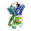 1amfS S: Starting model for refinement |
|---|---|
| Similar structure data |


-Links
 PDBj
PDBj- Assembly
| Deposited unit | 
| |||||||||
|---|---|---|---|---|---|---|---|---|---|---|
| 1 | 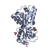
| |||||||||
| 2 | 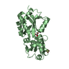
| |||||||||
| 3 | 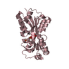
| |||||||||
| 4 | 
| |||||||||
| 5 | 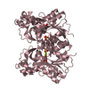
| |||||||||
| Unit cell |
| |||||||||
| Components on special symmetry positions |
|
-Components
| #1: Protein | Mass: 26696.189 Da / Num. of mol.: 3 / Fragment: residues 1-234 Source method: isolated from a genetically manipulated source Source: (gene. exp.) Xanthomonas axonopodis pv. citri (bacteria)Species: Xanthomonas axonopodis / Strain: 306 / Gene: modA / Plasmid: pET28a / Species (production host): Escherichia coli / Production host: #2: Chemical | ChemComp-SO4 /   Mass: 96.063 Da / Num. of mol.: 4 / Source method: obtained synthetically / Formula: SO4 Mass: 96.063 Da / Num. of mol.: 4 / Source method: obtained synthetically / Formula: SO4#3: Chemical |   Mass: 159.938 Da / Num. of mol.: 3 / Source method: obtained synthetically / Formula: MoO4 Mass: 159.938 Da / Num. of mol.: 3 / Source method: obtained synthetically / Formula: MoO4#4: Water | ChemComp-HOH / |  Mass: 18.015 Da / Num. of mol.: 829 / Source method: isolated from a natural source / Formula: H2O Mass: 18.015 Da / Num. of mol.: 829 / Source method: isolated from a natural source / Formula: H2OSequence details | THE RESIDUE IS LEU AND IS PROBABLY A REAL VARIANT PRESENT IN NATURE. | |
|---|
-Experimental details
-Experiment
| Experiment | Method: X-RAY DIFFRACTION / Number of used crystals: 1 |
|---|
- Sample preparation
| Crystal | Density Matthews: 2.05 Å3/Da / Density % sol: 40.04 % |
|---|---|
| Crystal grow | Temperature: 291 K / Method: vapor diffusion, sitting drop / pH: 8.5 Details: 0.2M lithium sulfate, 0.1M Tris-HCl, 30% PEG 4000 , pH 8.5, VAPOR DIFFUSION, SITTING DROP, temperature 291K |
-Data collection
| Diffraction | Mean temperature: 100 K |
|---|---|
| Diffraction source | Source: SYNCHROTRON / Site: LNLS  / Beamline: D03B-MX1 / Wavelength: 1.421 Å / Beamline: D03B-MX1 / Wavelength: 1.421 Å |
| Detector | Type: MAR CCD 165 mm / Detector: CCD / Date: Jan 13, 2005 / Details: mirror |
| Radiation | Monochromator: Si crystal / Protocol: SINGLE WAVELENGTH / Monochromatic (M) / Laue (L): M / Scattering type: x-ray |
| Radiation wavelength | Wavelength: 1.421 Å / Relative weight: 1 |
| Reflection | Resolution: 1.7→50 Å / Num. all: 72786 / Num. obs: 72786 / % possible obs: 99.8 % / Observed criterion σ(F): 0 / Observed criterion σ(I): 0 / Redundancy: 5.7 % / Rsym value: 0.062 / Net I/σ(I): 26.6 |
| Reflection shell | Resolution: 1.7→1.76 Å / Redundancy: 5.7 % / Mean I/σ(I) obs: 3.3 / Rsym value: 0.415 / % possible all: 99.3 |
- Processing
| Software |
| ||||||||||||||||||||||||||||||||||||||||||||||||||||||||||||||||||||||||||||||||||||||||||||||||||||||||||||||||||||||||||||||||||||||||||||||||||||||||||||||||||||||||||
|---|---|---|---|---|---|---|---|---|---|---|---|---|---|---|---|---|---|---|---|---|---|---|---|---|---|---|---|---|---|---|---|---|---|---|---|---|---|---|---|---|---|---|---|---|---|---|---|---|---|---|---|---|---|---|---|---|---|---|---|---|---|---|---|---|---|---|---|---|---|---|---|---|---|---|---|---|---|---|---|---|---|---|---|---|---|---|---|---|---|---|---|---|---|---|---|---|---|---|---|---|---|---|---|---|---|---|---|---|---|---|---|---|---|---|---|---|---|---|---|---|---|---|---|---|---|---|---|---|---|---|---|---|---|---|---|---|---|---|---|---|---|---|---|---|---|---|---|---|---|---|---|---|---|---|---|---|---|---|---|---|---|---|---|---|---|---|---|---|---|---|---|
| Refinement | Method to determine structure: MOLECULAR REPLACEMENT Starting model: PDB ENTRY 1AMF Resolution: 1.7→30.07 Å / Cor.coef. Fo:Fc: 0.963 / Cor.coef. Fo:Fc free: 0.943 / SU B: 3.472 / SU ML: 0.061 / TLS residual ADP flag: LIKELY RESIDUAL / Cross valid method: THROUGHOUT / ESU R: 0.104 / ESU R Free: 0.102 / Stereochemistry target values: MAXIMUM LIKELIHOOD Details: HYDROGENS HAVE BEEN ADDED IN THE RIDING POSITIONS.; Although waters 481 and 659 are about 0.02 Ang outside the limit of 0.15 Ang stated in the remark 375, they are considered by the authors ...Details: HYDROGENS HAVE BEEN ADDED IN THE RIDING POSITIONS.; Although waters 481 and 659 are about 0.02 Ang outside the limit of 0.15 Ang stated in the remark 375, they are considered by the authors to be in special positions.
| ||||||||||||||||||||||||||||||||||||||||||||||||||||||||||||||||||||||||||||||||||||||||||||||||||||||||||||||||||||||||||||||||||||||||||||||||||||||||||||||||||||||||||
| Solvent computation | Ion probe radii: 0.8 Å / Shrinkage radii: 0.8 Å / VDW probe radii: 1.4 Å / Solvent model: BABINET MODEL WITH MASK | ||||||||||||||||||||||||||||||||||||||||||||||||||||||||||||||||||||||||||||||||||||||||||||||||||||||||||||||||||||||||||||||||||||||||||||||||||||||||||||||||||||||||||
| Displacement parameters | Biso mean: 25.879 Å2
| ||||||||||||||||||||||||||||||||||||||||||||||||||||||||||||||||||||||||||||||||||||||||||||||||||||||||||||||||||||||||||||||||||||||||||||||||||||||||||||||||||||||||||
| Refinement step | Cycle: LAST / Resolution: 1.7→30.07 Å
| ||||||||||||||||||||||||||||||||||||||||||||||||||||||||||||||||||||||||||||||||||||||||||||||||||||||||||||||||||||||||||||||||||||||||||||||||||||||||||||||||||||||||||
| Refine LS restraints |
| ||||||||||||||||||||||||||||||||||||||||||||||||||||||||||||||||||||||||||||||||||||||||||||||||||||||||||||||||||||||||||||||||||||||||||||||||||||||||||||||||||||||||||
| LS refinement shell | Resolution: 1.697→1.741 Å / Total num. of bins used: 20
| ||||||||||||||||||||||||||||||||||||||||||||||||||||||||||||||||||||||||||||||||||||||||||||||||||||||||||||||||||||||||||||||||||||||||||||||||||||||||||||||||||||||||||
| Refinement TLS params. | Method: refined / Refine-ID: X-RAY DIFFRACTION
| ||||||||||||||||||||||||||||||||||||||||||||||||||||||||||||||||||||||||||||||||||||||||||||||||||||||||||||||||||||||||||||||||||||||||||||||||||||||||||||||||||||||||||
| Refinement TLS group |
|
