 Movie
Movie Controller


+ Open data

- Basic information
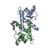
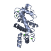
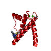
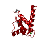
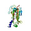
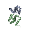
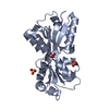
| Entry | Database: PDB / ID: 1mho | ||||||
|---|---|---|---|---|---|---|---|
| Title | THE 2.0 A STRUCTURE OF HOLO S100B FROM BOVINE BRAIN | ||||||
 Components Components | S-100 PROTEIN | ||||||
 Keywords Keywords | CALCIUM-BINDING / METAL-BINDING | ||||||
| Function / homology |  Function and homology information Function and homology informationAdvanced glycosylation endproduct receptor signaling / TRAF6 mediated NF-kB activation / TAK1-dependent IKK and NF-kappa-B activation / adaptive thermogenesis / kinase inhibitor activity / sympathetic neuron projection extension / positive regulation of complement activation / RAGE receptor binding / negative regulation of monocyte chemotactic protein-1 production / S100 protein binding ...Advanced glycosylation endproduct receptor signaling / TRAF6 mediated NF-kB activation / TAK1-dependent IKK and NF-kappa-B activation / adaptive thermogenesis / kinase inhibitor activity / sympathetic neuron projection extension / positive regulation of complement activation / RAGE receptor binding / negative regulation of monocyte chemotactic protein-1 production / S100 protein binding / phosphorylation / regulation of neuronal synaptic plasticity / ruffle / positive regulation of neuron differentiation / axonogenesis / astrocyte activation / tau protein binding / memory / calcium-dependent protein binding / regulation of translation / learning or memory / positive regulation of canonical NF-kappaB signal transduction / cell adhesion / ciliary basal body / neuronal cell body / calcium ion binding / positive regulation of cell population proliferation / perinuclear region of cytoplasm / protein homodimerization activity / : / extracellular region / zinc ion binding / nucleoplasm / nucleus / cytoplasm / cytosol Similarity search - Function | ||||||
| Biological species |  | ||||||
| Method |  X-RAY DIFFRACTION / MOLECULAR REPLACEMENT / Resolution: 2 Å X-RAY DIFFRACTION / MOLECULAR REPLACEMENT / Resolution: 2 Å | ||||||
 Authors Authors | Matsumura, H. / Shiba, T. / Inoue, T. / Harada, S. / Yasushi, K.A.I. | ||||||
 Citation Citation | Journal: Structure / Year: 1998 Title: A novel mode of target recognition suggested by the 2.0 A structure of holo S100B from bovine brain. Authors: Matsumura, H. / Shiba, T. / Inoue, T. / Harada, S. / Kai, Y. #1: Journal: J.Biol.Chem. / Year: 1986Title: The Refined Structure of Vitamin D-Dependent Calcium-Binding Protein from Bovine Intestine. Molecular Details, Ion Binding, and Implications for the Structure of Other Calcium-Binding Proteins Authors: Szebenyi, D.M. / Moffat, K. | ||||||
| History |
|
- Structure visualization
| Structure viewer | Molecule: MolmilJmol/JSmol |
|---|
- Downloads & links
-Download
| PDBx/mmCIF format | 1mho.cif.gz | 31.2 KB | Display | PDBx/mmCIF format |
|---|---|---|---|---|
| PDB format | pdb1mho.ent.gz | 20.7 KB | Display | PDB format |
| PDBx/mmJSON format | 1mho.json.gz | Tree view | PDBx/mmJSON format | |
| Others |  Other downloads Other downloads |
-Validation report
| Arichive directory | https://data.pdbj.org/pub/pdb/validation_reports/mh/1mhoftp://data.pdbj.org/pub/pdb/validation_reports/mh/1mho | HTTPS FTP |
|---|
-Related structure data
| Similar structure data |
|---|


-Links
 PDBj
PDBj- Assembly
| Deposited unit | 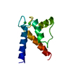
| ||||||||
|---|---|---|---|---|---|---|---|---|---|
| 1 | 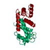
| ||||||||
| Unit cell |
|
-Components
| #1: Protein | Mass: 10154.402 Da / Num. of mol.: 1 / Fragment: BETA CHAIN / Source method: isolated from a natural source / Source: (natural) | ||
|---|---|---|---|
| #2: Chemical |   Mass: 40.078 Da / Num. of mol.: 2 / Source method: obtained synthetically / Formula: Ca Mass: 40.078 Da / Num. of mol.: 2 / Source method: obtained synthetically / Formula: Ca#3: Water | ChemComp-HOH / |  Mass: 18.015 Da / Num. of mol.: 59 / Source method: isolated from a natural source / Formula: H2O Mass: 18.015 Da / Num. of mol.: 59 / Source method: isolated from a natural source / Formula: H2O |
-Experimental details
-Experiment
| Experiment | Method: X-RAY DIFFRACTION / Number of used crystals: 1 |
|---|
- Sample preparation
| Crystal | Density Matthews: 2.24 Å3/Da / Density % sol: 45 % | ||||||||||||||||||||||||||||||
|---|---|---|---|---|---|---|---|---|---|---|---|---|---|---|---|---|---|---|---|---|---|---|---|---|---|---|---|---|---|---|---|
| Crystal grow | Method: vapor diffusion, hanging drop / pH: 8 Details: HANGING-DROP VAPOR DIFFUSION METHOD, pH 8.0, vapor diffusion - hanging drop | ||||||||||||||||||||||||||||||
| Crystal | *PLUS | ||||||||||||||||||||||||||||||
| Crystal grow | *PLUS Temperature: 20 ℃ / pH: 6.1 / Method: vapor diffusion, hanging drop | ||||||||||||||||||||||||||||||
| Components of the solutions | *PLUS
|
-Data collection
| Diffraction | Mean temperature: 298 K |
|---|---|
| Diffraction source | Source: ROTATING ANODE / Type: RIGAKU RUH3R / Wavelength: 1.5418 |
| Detector | Type: RIGAKU / Detector: IMAGE PLATE / Date: Oct 2, 1995 |
| Radiation | Monochromator: GRAPHITE(002) / Monochromatic (M) / Laue (L): M / Scattering type: x-ray |
| Radiation wavelength | Wavelength: 1.5418 Å / Relative weight: 1 |
| Reflection | Resolution: 2→30 Å / Num. obs: 6282 / % possible obs: 93.8 % / Observed criterion σ(I): 1 / Redundancy: 2.7 % / Biso Wilson estimate: 27.73 Å2 / Rmerge(I) obs: 0.061 / Net I/σ(I): 7 |
| Reflection shell | Resolution: 2→2.15 Å / Rmerge(I) obs: 0.171 / % possible all: 91.3 |
| Reflection | *PLUS Num. measured all: 17098 |
- Processing
| Software |
| ||||||||||||||||||||||||||||||||||||||||||||||||||||||||||||||||||||||||||||||||||||
|---|---|---|---|---|---|---|---|---|---|---|---|---|---|---|---|---|---|---|---|---|---|---|---|---|---|---|---|---|---|---|---|---|---|---|---|---|---|---|---|---|---|---|---|---|---|---|---|---|---|---|---|---|---|---|---|---|---|---|---|---|---|---|---|---|---|---|---|---|---|---|---|---|---|---|---|---|---|---|---|---|---|---|---|---|---|
| Refinement | Method to determine structure: MOLECULAR REPLACEMENT Starting model: VITAMIN D-DEPENDENT CALCIUM-BINDING PROTEIN FROM BOVINE INTESTINE Resolution: 2→10 Å
| ||||||||||||||||||||||||||||||||||||||||||||||||||||||||||||||||||||||||||||||||||||
| Displacement parameters | Biso mean: 31.69 Å2 | ||||||||||||||||||||||||||||||||||||||||||||||||||||||||||||||||||||||||||||||||||||
| Refinement step | Cycle: LAST / Resolution: 2→10 Å
| ||||||||||||||||||||||||||||||||||||||||||||||||||||||||||||||||||||||||||||||||||||
| Refine LS restraints |
| ||||||||||||||||||||||||||||||||||||||||||||||||||||||||||||||||||||||||||||||||||||
| Software | *PLUS Name: REFMAC / Classification: refinement | ||||||||||||||||||||||||||||||||||||||||||||||||||||||||||||||||||||||||||||||||||||
| Refinement | *PLUS Rfactor obs: 0.194 | ||||||||||||||||||||||||||||||||||||||||||||||||||||||||||||||||||||||||||||||||||||
| Solvent computation | *PLUS | ||||||||||||||||||||||||||||||||||||||||||||||||||||||||||||||||||||||||||||||||||||
| Displacement parameters | *PLUS |
