 Movie
Movie Controller

[English] 日本語
 Yorodumi
- PDB-1amf: CRYSTAL STRUCTURE OF MODA, A MOLYBDATE TRANSPORT PROTEIN, COMPLEX... -
+ Open data

- Basic information
| Entry | Database: PDB / ID: 1amf | ||||||
|---|---|---|---|---|---|---|---|
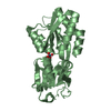
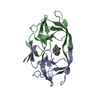
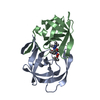
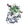
| Title | CRYSTAL STRUCTURE OF MODA, A MOLYBDATE TRANSPORT PROTEIN, COMPLEXED WITH MOLYBDATE | ||||||
 Components Components | MOLYBDATE TRANSPORT PROTEIN MODA | ||||||
 Keywords Keywords | BINDING PROTEIN / MOLYBDATE TRANSPORT PROTEIN / MOLYBDATE / PERIPLASMIC | ||||||
| Function / homology |  Function and homology information Function and homology informationresponse to chromate / tungstate ion transport / ABC-type molybdate transporter activity / tungstate binding / molybdate ion binding / molybdate ion transport / molybdenum ion binding / ATP-binding cassette (ABC) transporter complex, substrate-binding subunit-containing / outer membrane-bounded periplasmic space / membrane Similarity search - Function | ||||||
| Biological species |  | ||||||
| Method |  X-RAY DIFFRACTION / SIRAS / Resolution: 1.75 Å X-RAY DIFFRACTION / SIRAS / Resolution: 1.75 Å | ||||||
 Authors Authors | Hu, Y. / Rech, S. / Gunsalus, R.P. / Rees, D.C. | ||||||
 Citation Citation | Journal: Nat.Struct.Biol. / Year: 1997 Title: Crystal structure of the molybdate binding protein ModA. Authors: Hu, Y. / Rech, S. / Gunsalus, R.P. / Rees, D.C. #1: Journal: J.Biol.Chem. / Year: 1996Title: Properties of the Periplasmic Moda Molybdate-Binding Protein of Escherichia Coli Authors: Rech, S. / Wolin, C. / Gunsalus, R.P. | ||||||
| History |
|
- Structure visualization
| Structure viewer | Molecule: MolmilJmol/JSmol |
|---|
- Downloads & links
-Download
| PDBx/mmCIF format | 1amf.cif.gz | 65.8 KB | Display | PDBx/mmCIF format |
|---|---|---|---|---|
| PDB format | pdb1amf.ent.gz | 50 KB | Display | PDB format |
| PDBx/mmJSON format | 1amf.json.gz | Tree view | PDBx/mmJSON format | |
| Others |  Other downloads Other downloads |
-Validation report
| Arichive directory | https://data.pdbj.org/pub/pdb/validation_reports/am/1amfftp://data.pdbj.org/pub/pdb/validation_reports/am/1amf | HTTPS FTP |
|---|
-Related structure data


-Links
 PDBj
PDBj- Assembly
| Deposited unit | 
| ||||||||
|---|---|---|---|---|---|---|---|---|---|
| 1 |
| ||||||||
| Unit cell |
|
-Components
| #1: Protein | Mass: 24948.264 Da / Num. of mol.: 1 / Fragment: N-DOMAIN, C-DOMAIN / Source method: isolated from a natural source Details: MOLYBDATE ANION IS SEQUESTERED BETWEEN N-AND C-DOMAINS Source: (natural) |
|---|---|
| #2: Chemical | ChemComp-MOO /   Mass: 159.938 Da / Num. of mol.: 1 / Source method: obtained synthetically / Formula: MoO4 Mass: 159.938 Da / Num. of mol.: 1 / Source method: obtained synthetically / Formula: MoO4 |
| #3: Water | ChemComp-HOH /  Mass: 18.015 Da / Num. of mol.: 76 / Source method: isolated from a natural source / Formula: H2O Mass: 18.015 Da / Num. of mol.: 76 / Source method: isolated from a natural source / Formula: H2O |
-Experimental details
-Experiment
| Experiment | Method: X-RAY DIFFRACTION / Number of used crystals: 1 |
|---|
- Sample preparation
| Crystal | Density Matthews: 3.1 Å3/Da / Density % sol: 61.73 % | ||||||||||||||||||||||||||||||||||||||||||
|---|---|---|---|---|---|---|---|---|---|---|---|---|---|---|---|---|---|---|---|---|---|---|---|---|---|---|---|---|---|---|---|---|---|---|---|---|---|---|---|---|---|---|---|
| Crystal grow | pH: 4.7 Details: PROTEIN WAS CRYSTALLIZED FROM 27.5% PEG 8000, 0.1M SODIUM ACETATE, PH 4.7, AND 2MM SODIUM MOLYBDATE | ||||||||||||||||||||||||||||||||||||||||||
| Crystal grow | *PLUS Method: vapor diffusion, hanging drop | ||||||||||||||||||||||||||||||||||||||||||
| Components of the solutions | *PLUS
|
-Data collection
| Diffraction | Mean temperature: 300 K |
|---|---|
| Diffraction source | Source: ROTATING ANODE / Type: RIGAKU RUH2R / Wavelength: 1.5418 |
| Detector | Type: RIGAKU RAXIS / Detector: IMAGE PLATE / Date: Aug 1, 1996 |
| Radiation | Monochromatic (M) / Laue (L): M / Scattering type: x-ray |
| Radiation wavelength | Wavelength: 1.5418 Å / Relative weight: 1 |
| Reflection | Resolution: 1.75→30 Å / Num. obs: 30575 / % possible obs: 93.2 % / Redundancy: 3.7 % / Biso Wilson estimate: 24.9 Å2 / Rsym value: 0.055 / Net I/σ(I): 21 |
| Reflection shell | Resolution: 1.75→1.81 Å / Redundancy: 1.4 % / Mean I/σ(I) obs: 1.9 / Rsym value: 0.244 / % possible all: 60.8 |
| Reflection | *PLUS Num. measured all: 112116 / Rmerge(I) obs: 0.055 |
| Reflection shell | *PLUS % possible obs: 60.8 % / Rmerge(I) obs: 0.244 |
- Processing
| Software |
| ||||||||||||||||||||||||||||||||||||||||||||||||||||||||||||
|---|---|---|---|---|---|---|---|---|---|---|---|---|---|---|---|---|---|---|---|---|---|---|---|---|---|---|---|---|---|---|---|---|---|---|---|---|---|---|---|---|---|---|---|---|---|---|---|---|---|---|---|---|---|---|---|---|---|---|---|---|---|
| Refinement | Method to determine structure: SIRAS / Resolution: 1.75→30 Å
| ||||||||||||||||||||||||||||||||||||||||||||||||||||||||||||
| Displacement parameters | Biso mean: 28.6 Å2 | ||||||||||||||||||||||||||||||||||||||||||||||||||||||||||||
| Refinement step | Cycle: LAST / Resolution: 1.75→30 Å
| ||||||||||||||||||||||||||||||||||||||||||||||||||||||||||||
| Refine LS restraints |
| ||||||||||||||||||||||||||||||||||||||||||||||||||||||||||||
| Xplor file |
| ||||||||||||||||||||||||||||||||||||||||||||||||||||||||||||
| Software | *PLUS Name: X-PLOR / Version: 4 / Classification: refinement | ||||||||||||||||||||||||||||||||||||||||||||||||||||||||||||
| Refinement | *PLUS | ||||||||||||||||||||||||||||||||||||||||||||||||||||||||||||
| Solvent computation | *PLUS | ||||||||||||||||||||||||||||||||||||||||||||||||||||||||||||
| Displacement parameters | *PLUS | ||||||||||||||||||||||||||||||||||||||||||||||||||||||||||||
| Refine LS restraints | *PLUS
|
