 Movie
Movie Controller

[English] 日本語
 Yorodumi
- PDB-5sv6: Crystal structure of MxaJ from Methlophaga aminisulfidivorans MPT -
+ Open data

- Basic information
| Entry | Database: PDB / ID: 5sv6 | ||||||
|---|---|---|---|---|---|---|---|
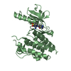
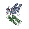
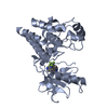
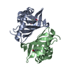
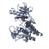
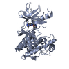
| Title | Crystal structure of MxaJ from Methlophaga aminisulfidivorans MPT | ||||||
 Components Components | Extracellular solute-binding protein, family 3 | ||||||
 Keywords Keywords | UNKNOWN FUNCTION / MxaJ / Methlophaga aminisulfidivorans / MDH / PBP | ||||||
| Function / homology | methanol catabolic process / Methanol oxidation system protein MoxJ / Bacterial extracellular solute-binding proteins, family 3 / Solute-binding protein family 3/N-terminal domain of MltF / Bacterial periplasmic substrate-binding proteins / periplasmic space / BROMIDE ION / Extracellular solute-binding protein, family 3 Function and homology information Function and homology information | ||||||
| Biological species |  Methylophaga aminisulfidivorans MP (bacteria) Methylophaga aminisulfidivorans MP (bacteria) | ||||||
| Method |  X-RAY DIFFRACTION / SYNCHROTRON / SAD / Resolution: 1.92 Å X-RAY DIFFRACTION / SYNCHROTRON / SAD / Resolution: 1.92 Å | ||||||
 Authors Authors | Choi, J.M. / Lee, S.H. | ||||||
 Citation Citation | Journal: Proteins / Year: 2017 Title: MxaJ structure reveals a periplasmic binding protein-like architecture with unique secondary structural elements Authors: Choi, J.M. / Cao, T.P. / Kim, S.W. / Lee, K.H. / Lee, S.H. | ||||||
| History |
|
- Structure visualization
| Structure viewer | Molecule: MolmilJmol/JSmol |
|---|
- Downloads & links
-Download
| PDBx/mmCIF format | 5sv6.cif.gz | 70.5 KB | Display | PDBx/mmCIF format |
|---|---|---|---|---|
| PDB format | pdb5sv6.ent.gz | 50.1 KB | Display | PDB format |
| PDBx/mmJSON format | 5sv6.json.gz | Tree view | PDBx/mmJSON format | |
| Others |  Other downloads Other downloads |
-Validation report
| Arichive directory | https://data.pdbj.org/pub/pdb/validation_reports/sv/5sv6ftp://data.pdbj.org/pub/pdb/validation_reports/sv/5sv6 | HTTPS FTP |
|---|
-Related structure data
| Similar structure data |
|---|


-Links
 PDBj
PDBj

- Assembly
| Deposited unit | 
| ||||||||
|---|---|---|---|---|---|---|---|---|---|
| 1 |
| ||||||||
| Unit cell |
|
-Components
| #1: Protein | Mass: 32839.879 Da / Num. of mol.: 1 / Fragment: UNP residues 12-281 Source method: isolated from a genetically manipulated source Source: (gene. exp.) Methylophaga aminisulfidivorans MP (bacteria)Strain: MP / Gene: mxaJ / Plasmid: pET28a(+) / Production host: |
|---|---|
| #2: Chemical | ChemComp-BR /   Mass: 79.904 Da / Num. of mol.: 1 / Source method: obtained synthetically / Formula: Br Mass: 79.904 Da / Num. of mol.: 1 / Source method: obtained synthetically / Formula: Br |
| #3: Water | ChemComp-HOH /  Mass: 18.015 Da / Num. of mol.: 285 / Source method: isolated from a natural source / Formula: H2O Mass: 18.015 Da / Num. of mol.: 285 / Source method: isolated from a natural source / Formula: H2O |
| Has protein modification | Y |
-Experimental details
-Experiment
| Experiment | Method: X-RAY DIFFRACTION / Number of used crystals: 1 |
|---|
- Sample preparation
| Crystal | Density Matthews: 1.79 Å3/Da / Density % sol: 31.23 % |
|---|---|
| Crystal grow | Temperature: 293 K / Method: vapor diffusion, hanging drop / pH: 8.5 Details: 0.2M sodium bromide, 20%(w/v) PEG 3350, 10%(w/v) glycerol, 0.1M Tris HCl |
-Data collection
| Diffraction | Mean temperature: 100 K |
|---|---|
| Diffraction source | Source: SYNCHROTRON / Site: PAL/PLS  / Beamline: 5C (4A) / Wavelength: 0.979 Å / Beamline: 5C (4A) / Wavelength: 0.979 Å |
| Detector | Type: ADSC QUANTUM 315r / Detector: CCD / Date: Dec 1, 2010 |
| Radiation | Monochromator: DCM Si(111) crystal / Protocol: MAD / Monochromatic (M) / Laue (L): M / Scattering type: x-ray |
| Radiation wavelength | Wavelength: 0.979 Å / Relative weight: 1 |
| Reflection | Resolution: 1.92→50 Å / Num. obs: 18684 / % possible obs: 99.9 % / Observed criterion σ(F): 0 / Observed criterion σ(I): -3 / Redundancy: 13.9 % / Rmerge(I) obs: 0.071 / Net I/σ(I): 0.69 |
| Reflection shell | Resolution: 1.92→1.95 Å / Redundancy: 13.3 % / Rmerge(I) obs: 0.254 / Mean I/σ(I) obs: 16.049 / % possible all: 100 |
- Processing
| Software |
| ||||||||||||||||||||||||||||||||||||||||||||||||||||||||||||||||||||||||||||||||||||||||||||||||||
|---|---|---|---|---|---|---|---|---|---|---|---|---|---|---|---|---|---|---|---|---|---|---|---|---|---|---|---|---|---|---|---|---|---|---|---|---|---|---|---|---|---|---|---|---|---|---|---|---|---|---|---|---|---|---|---|---|---|---|---|---|---|---|---|---|---|---|---|---|---|---|---|---|---|---|---|---|---|---|---|---|---|---|---|---|---|---|---|---|---|---|---|---|---|---|---|---|---|---|---|
| Refinement | Method to determine structure: SAD / Resolution: 1.92→29.728 Å / SU ML: 0.19 / Cross valid method: THROUGHOUT / σ(F): 0.33 / Phase error: 21.2 / Stereochemistry target values: ML
| ||||||||||||||||||||||||||||||||||||||||||||||||||||||||||||||||||||||||||||||||||||||||||||||||||
| Solvent computation | Shrinkage radii: 0.9 Å / VDW probe radii: 1.11 Å / Solvent model: FLAT BULK SOLVENT MODEL | ||||||||||||||||||||||||||||||||||||||||||||||||||||||||||||||||||||||||||||||||||||||||||||||||||
| Refinement step | Cycle: LAST / Resolution: 1.92→29.728 Å
| ||||||||||||||||||||||||||||||||||||||||||||||||||||||||||||||||||||||||||||||||||||||||||||||||||
| Refine LS restraints |
| ||||||||||||||||||||||||||||||||||||||||||||||||||||||||||||||||||||||||||||||||||||||||||||||||||
| LS refinement shell |
|
