| Entry | Database: PDB / ID: 6p3d
|
|---|
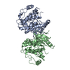
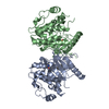
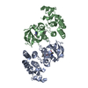
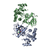
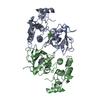
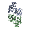
| Title | The co-crystal structure of BRAF(V600E) with ponatinib |
|---|
 Components Components | Serine/threonine-protein kinase B-raf |
|---|
 Keywords Keywords | TRANSFERASE/TRANSFERASE INHIBITOR / kinase / inhibitor / cancer / melanoma / TRANSFERASE-TRANSFERASE INHIBITOR complex / ONCOPROTEIN |
|---|
| Function / homology |  Function and homology information Function and homology information
CD4-positive, alpha-beta T cell differentiation / positive regulation of axon regeneration / CD4-positive or CD8-positive, alpha-beta T cell lineage commitment / negative regulation of synaptic vesicle exocytosis / Signalling to p38 via RIT and RIN / head morphogenesis / myeloid progenitor cell differentiation / ARMS-mediated activation / endothelial cell apoptotic process / SHOC2 M1731 mutant abolishes MRAS complex function ...CD4-positive, alpha-beta T cell differentiation / positive regulation of axon regeneration / CD4-positive or CD8-positive, alpha-beta T cell lineage commitment / negative regulation of synaptic vesicle exocytosis / Signalling to p38 via RIT and RIN / head morphogenesis / myeloid progenitor cell differentiation / ARMS-mediated activation / endothelial cell apoptotic process / SHOC2 M1731 mutant abolishes MRAS complex function / Gain-of-function MRAS complexes activate RAF signaling / negative regulation of fibroblast migration / positive regulation of D-glucose transmembrane transport / establishment of protein localization to membrane / regulation of T cell differentiation / positive regulation of axonogenesis / Negative feedback regulation of MAPK pathway / Frs2-mediated activation / stress fiber assembly / face development / MAP kinase kinase activity / thyroid gland development / somatic stem cell population maintenance / synaptic vesicle exocytosis / positive regulation of peptidyl-serine phosphorylation / MAP kinase kinase kinase activity / negative regulation of endothelial cell apoptotic process / postsynaptic modulation of chemical synaptic transmission / ERK1 and ERK2 cascade / positive regulation of stress fiber assembly / positive regulation of substrate adhesion-dependent cell spreading / substrate adhesion-dependent cell spreading / cellular response to calcium ion / thymus development / animal organ morphogenesis / sperm end piece / RAF activation / Signaling by high-kinase activity BRAF mutants / Spry regulation of FGF signaling / MAP2K and MAPK activation / visual learning / cellular response to xenobiotic stimulus / epidermal growth factor receptor signaling pathway / centriolar satellite / Signaling by RAF1 mutants / Signaling by moderate kinase activity BRAF mutants / Paradoxical activation of RAF signaling by kinase inactive BRAF / Signaling downstream of RAS mutants / Negative regulation of MAPK pathway / Signaling by BRAF and RAF1 fusions / long-term synaptic potentiation / T cell differentiation in thymus / MAPK cascade / regulation of cell population proliferation / T cell receptor signaling pathway / presynapse / sperm principal piece / cell body / scaffold protein binding / sperm midpiece / protein phosphorylation / negative regulation of neuron apoptotic process / protein kinase activity / positive regulation of ERK1 and ERK2 cascade / non-specific serine/threonine protein kinase / neuron projection / postsynapse / cilium / ciliary basal body / protein serine kinase activity / protein serine/threonine kinase activity / calcium ion binding / positive regulation of gene expression / negative regulation of apoptotic process / glutamatergic synapse / mitochondrion / zinc ion binding / ATP binding / identical protein binding / nucleus / plasma membrane / cytoplasm / cytosolSimilarity search - Function Raf-like Ras-binding domain / Raf-like Ras-binding / Ras-binding domain (RBD) profile. / Raf-like Ras-binding domain / Diacylglycerol/phorbol-ester binding / Phorbol esters/diacylglycerol binding domain (C1 domain) / : / Zinc finger phorbol-ester/DAG-type signature. / Zinc finger phorbol-ester/DAG-type profile. / Protein kinase C conserved region 1 (C1) domains (Cysteine-rich domains) ...Raf-like Ras-binding domain / Raf-like Ras-binding / Ras-binding domain (RBD) profile. / Raf-like Ras-binding domain / Diacylglycerol/phorbol-ester binding / Phorbol esters/diacylglycerol binding domain (C1 domain) / : / Zinc finger phorbol-ester/DAG-type signature. / Zinc finger phorbol-ester/DAG-type profile. / Protein kinase C conserved region 1 (C1) domains (Cysteine-rich domains) / Protein kinase C-like, phorbol ester/diacylglycerol-binding domain / C1-like domain superfamily / Protein tyrosine and serine/threonine kinase / Serine-threonine/tyrosine-protein kinase, catalytic domain / Ubiquitin-like domain superfamily / Transferase(Phosphotransferase) domain 1 / Transferase(Phosphotransferase); domain 1 / Serine/threonine-protein kinase, active site / Serine/Threonine protein kinases active-site signature. / Serine/Threonine protein kinases, catalytic domain / Protein kinase, ATP binding site / Protein kinases ATP-binding region signature. / Protein kinase domain profile. / Protein kinase domain / Protein kinase-like domain superfamily / Orthogonal Bundle / Mainly AlphaSimilarity search - Domain/homology |
|---|
| Biological species |  Homo sapiens (human) Homo sapiens (human) |
|---|
| Method |  X-RAY DIFFRACTION / SYNCHROTRON / MOLECULAR REPLACEMENT / Resolution: 2.11 Å X-RAY DIFFRACTION / SYNCHROTRON / MOLECULAR REPLACEMENT / Resolution: 2.11 Å |
|---|
 Authors Authors | Agianian, B. / Gavathiotis, E. |
|---|
| Funding support |  United States, 1items United States, 1items | Organization | Grant number | Country |
|---|
| National Institutes of Health/National Cancer Institute (NIH/NCI) | 1R01CA178394 | United States |
|
|---|
 Citation Citation | Journal: Nat Commun / Year: 2020Title: Inhibitors of BRAF dimers using an allosteric site. Authors: Cotto-Rios, X.M. / Agianian, B. / Gitego, N. / Zacharioudakis, E. / Giricz, O. / Wu, Y. / Zou, Y. / Verma, A. / Poulikakos, P.I. / Gavathiotis, E. #1: Journal: Cancer Cell / Year: 2016Title: An Integrated Model of RAF Inhibitor Action Predicts Inhibitor Activity against Oncogenic BRAF Signaling. Authors: Karoulia, Z. / Wu, Y. / Ahmed, T.A. / Xin, Q. / Bollard, J. / Krepler, C. / Wu, X. / Zhang, C. / Bollag, G. / Herlyn, M. / Fagin, J.A. / Lujambio, A. / Gavathiotis, E. / Poulikakos, P.I. |
|---|
| History | | Deposition | May 23, 2019 | Deposition site: RCSB / Processing site: RCSB |
|---|
| Revision 1.0 | Sep 23, 2020 | Provider: repository / Type: Initial release |
|---|
| Revision 1.1 | Mar 13, 2024 | Group: Data collection / Database references / Category: chem_comp_atom / chem_comp_bond / database_2
Item: _database_2.pdbx_DOI / _database_2.pdbx_database_accession |
|---|
|
|---|
 Movie
Movie Controller


 Open data
 Basic information
 Structure visualization
 Downloads & links
 Other downloads


 PDBj
PDBj


 Assembly


 Mass: 532.559 Da / Num. of mol.: 1 / Source method: obtained synthetically / Formula: C29H27F3N6O / Feature type: SUBJECT OF INVESTIGATION / Comment: medication*YM
 Mass: 96.063 Da / Num. of mol.: 8 / Source method: obtained synthetically / Formula: SO4
 Mass: 18.038 Da / Num. of mol.: 1 / Source method: obtained synthetically / Formula: H4N
 Mass: 62.068 Da / Num. of mol.: 2 / Source method: obtained synthetically / Formula: C2H6O2
 Sample preparation
 Processing