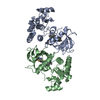
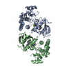
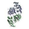
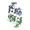
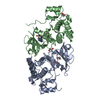
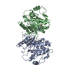
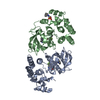
Entry Database : PDB / ID : 6p7gTitle The co-crystal structure of BRAF(V600E) with PHI1 Serine/threonine-protein kinase B-raf Keywords / / / / / / Function / homology Function Domain/homology Component
/ / / / / / / / / / / / / / / / / / / / / / / / / / / / / / / / / / / / / / / / / / / / / / / / / / / / / / / / / / / / / / / / / / / / / / / / / / / / / / / / / / / / / / / / / / / / / / / / / / / / / / / / / / / / / / / / / / / / / / / / / / / / / / Biological species Homo sapiens (human)Method / / / / Resolution : 2.65 Å Authors Agianian, B. / Gavathiotis, E. Funding support Organization Grant number Country National Institutes of Health/National Cancer Institute (NIH/NCI) R01CA178394 National Institutes of Health/National Cancer Institute (NIH/NCI) R01CA204314 National Institutes of Health/National Cancer Institute (NIH/NCI) P30CA013330
Journal : Nat Commun / Year : 2020Title : Inhibitors of BRAF dimers using an allosteric site.
Authors :
Cotto-Rios, X.M. / Agianian, B. / Gitego, N. / Zacharioudakis, E. / Giricz, O. / Wu, Y. / Zou, Y. / Verma, A. / Poulikakos, P.I. / Gavathiotis, E. #1: Journal : Cancer Cell / Year : 2016Title : An Integrated Model of RAF Inhibitor Action Predicts Inhibitor Activity against Oncogenic BRAF Signaling.
Authors :
Karoulia, Z. / Wu, Y. / Ahmed, T.A. / Xin, Q. / Bollard, J. / Krepler, C. / Wu, X. / Zhang, C. / Bollag, G. / Herlyn, M. / Fagin, J.A. / Lujambio, A. / Gavathiotis, E. / Poulikakos, P.I. History Deposition Jun 5, 2019 Deposition site / Processing site Revision 1.0 Sep 23, 2020 Provider / Type Revision 1.1 Mar 13, 2024 Group Data collection / Database references ... Data collection / Database references / Refinement description / Structure summary Category chem_comp_atom / chem_comp_bond ... chem_comp_atom / chem_comp_bond / database_2 / struct_keywords / struct_ncs_dom_lim Item _database_2.pdbx_DOI / _database_2.pdbx_database_accession ... _database_2.pdbx_DOI / _database_2.pdbx_database_accession / _struct_keywords.pdbx_keywords / _struct_keywords.text / _struct_ncs_dom_lim.beg_auth_comp_id / _struct_ncs_dom_lim.beg_label_asym_id / _struct_ncs_dom_lim.beg_label_comp_id / _struct_ncs_dom_lim.beg_label_seq_id / _struct_ncs_dom_lim.end_auth_comp_id / _struct_ncs_dom_lim.end_label_asym_id / _struct_ncs_dom_lim.end_label_comp_id / _struct_ncs_dom_lim.end_label_seq_id
Show all Show less
 Movie
Movie Controller


 Open data
 Basic information
 Components
 Keywords
 Function and homology information
 Homo sapiens (human)
 X-RAY DIFFRACTION /
  Authors
 United States, 3items
  Citation
 Structure visualization
 Downloads & links
 Other downloads
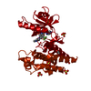

 PDBj
PDBj


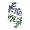
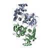
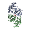
 Assembly
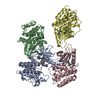


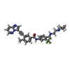
 Mass: 562.585 Da / Num. of mol.: 4
 Mass: 207.290 Da / Num. of mol.: 1 / Source method: obtained synthetically / Formula: C8H17NO3S / Comment: pH buffer*YM
 Mass: 18.015 Da / Num. of mol.: 50 / Source method: isolated from a natural source / Formula: H2O
 Sample preparation
 Processing