 Movie
Movie Controller

[English] 日本語
 Yorodumi
- PDB-1s50: X-ray structure of the GluR6 ligand binding core (S1S2A) in compl... -
+ Open data

- Basic information
| Entry | Database: PDB / ID: 1s50 | ||||||
|---|---|---|---|---|---|---|---|
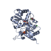
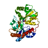
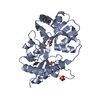
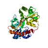
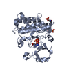
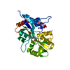
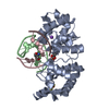
| Title | X-ray structure of the GluR6 ligand binding core (S1S2A) in complex with glutamate at 1.65 A resolution | ||||||
 Components Components | Glutamate Receptor 6 | ||||||
 Keywords Keywords | MEMBRANE PROTEIN | ||||||
| Function / homology |  Function and homology information Function and homology informationmossy fiber rosette / detection of cold stimulus involved in thermoception / Activation of Na-permeable kainate receptors / regulation of short-term neuronal synaptic plasticity / Activation of Ca-permeable Kainate Receptor / kainate selective glutamate receptor complex / ubiquitin conjugating enzyme binding / glutamate receptor activity / negative regulation of synaptic transmission, glutamatergic / regulation of JNK cascade ...mossy fiber rosette / detection of cold stimulus involved in thermoception / Activation of Na-permeable kainate receptors / regulation of short-term neuronal synaptic plasticity / Activation of Ca-permeable Kainate Receptor / kainate selective glutamate receptor complex / ubiquitin conjugating enzyme binding / glutamate receptor activity / negative regulation of synaptic transmission, glutamatergic / regulation of JNK cascade / inhibitory postsynaptic potential / receptor clustering / kainate selective glutamate receptor activity / extracellularly glutamate-gated ion channel activity / modulation of excitatory postsynaptic potential / ionotropic glutamate receptor complex / behavioral fear response / positive regulation of synaptic transmission / neuronal action potential / glutamate-gated receptor activity / glutamate-gated calcium ion channel activity / dendrite cytoplasm / ligand-gated monoatomic ion channel activity involved in regulation of presynaptic membrane potential / presynaptic modulation of chemical synaptic transmission / excitatory postsynaptic potential / hippocampal mossy fiber to CA3 synapse / SNARE binding / PDZ domain binding / regulation of long-term neuronal synaptic plasticity / synaptic transmission, glutamatergic / regulation of membrane potential / transmitter-gated monoatomic ion channel activity involved in regulation of postsynaptic membrane potential / intracellular protein transport / postsynaptic density membrane / modulation of chemical synaptic transmission / intracellular calcium ion homeostasis / terminal bouton / positive regulation of neuron apoptotic process / neuron apoptotic process / presynaptic membrane / scaffold protein binding / chemical synaptic transmission / negative regulation of neuron apoptotic process / perikaryon / postsynaptic membrane / postsynaptic density / axon / neuronal cell body / ubiquitin protein ligase binding / dendrite / synapse / glutamatergic synapse / membrane / identical protein binding / plasma membrane Similarity search - Function | ||||||
| Biological species |  | ||||||
| Method |  X-RAY DIFFRACTION / SYNCHROTRON / MOLECULAR REPLACEMENT / Resolution: 1.65 Å X-RAY DIFFRACTION / SYNCHROTRON / MOLECULAR REPLACEMENT / Resolution: 1.65 Å | ||||||
 Authors Authors | Mayer, M.L. | ||||||
 Citation Citation | Journal: Neuron / Year: 2005 Title: Crystal structures of the GluR5 and GluR6 ligand binding cores: Molecular mechanisms underlying kainate receptor selectivity Authors: Mayer, M.L. | ||||||
| History |
| ||||||
| Remark 999 | SEQUENCE THE FIRST GLY IS VECTOR ENCODED. THE NATIVE GLUR-5 IS A MEMBRANE PROTEIN. THE PROTEIN ... SEQUENCE THE FIRST GLY IS VECTOR ENCODED. THE NATIVE GLUR-5 IS A MEMBRANE PROTEIN. THE PROTEIN CRYSTALLIZED BY THE AUTHOR IS THE EXTRACELLULAR LIGAND BINDING DOMAIN OF GLUR-5. TRANSMEMBRANE REGIONS WERE GENETICALLY REMOVED AND REPLACED WITH A GLY-THR LINKER. THE SEQUENCE, AS A RESULT, MATCHES DISCONTINUOUSLY WITH THE REFERENCE DATABASE | ||||||
| Remark 300 | BIOMOLECULE: 1 THIS ENTRY CONTAINS THE CRYSTALLOGRAPHIC ASYMMETRIC UNIT WHICH CONSISTS OF 1 CHAIN. ... BIOMOLECULE: 1 THIS ENTRY CONTAINS THE CRYSTALLOGRAPHIC ASYMMETRIC UNIT WHICH CONSISTS OF 1 CHAIN. THE BIOLOGICAL UNIT IS BELIEVED TO BE A DIMER, BUT IN THIS CRYSTAL FORM THERE AREN'T ANY SYMMETRY OPERATIONS WHICH GENERATE THE DIMER. |
- Structure visualization
| Structure viewer | Molecule: MolmilJmol/JSmol |
|---|
- Downloads & links
-Download
| PDBx/mmCIF format | 1s50.cif.gz | 73.4 KB | Display | PDBx/mmCIF format |
|---|---|---|---|---|
| PDB format | pdb1s50.ent.gz | 52.9 KB | Display | PDB format |
| PDBx/mmJSON format | 1s50.json.gz | Tree view | PDBx/mmJSON format | |
| Others |  Other downloads Other downloads |
-Validation report
| Arichive directory | https://data.pdbj.org/pub/pdb/validation_reports/s5/1s50ftp://data.pdbj.org/pub/pdb/validation_reports/s5/1s50 | HTTPS FTP |
|---|
-Related structure data
| Related structure data |  1s7ySC  1s9tC  1sd3C  1tt1C  1txfC S: Starting model for refinement C: citing same article ( |
|---|---|
| Similar structure data |


-Links
 PDBj
PDBj


- Assembly
| Deposited unit | 
| ||||||||
|---|---|---|---|---|---|---|---|---|---|
| 1 |
| ||||||||
| Unit cell |
|
-Components
| #1: Protein | Mass: 29371.633 Da / Num. of mol.: 1 / Fragment: GluR6 ligand binding core Source method: isolated from a genetically manipulated source Source: (gene. exp.)  |
|---|---|
| #2: Chemical | ChemComp-GLU /   Type: L-peptide linking / Mass: 147.129 Da / Num. of mol.: 1 / Source method: obtained synthetically / Formula: C5H9NO4 Type: L-peptide linking / Mass: 147.129 Da / Num. of mol.: 1 / Source method: obtained synthetically / Formula: C5H9NO4 |
| #3: Water | ChemComp-HOH /  Mass: 18.015 Da / Num. of mol.: 359 / Source method: isolated from a natural source / Formula: H2O Mass: 18.015 Da / Num. of mol.: 359 / Source method: isolated from a natural source / Formula: H2O |
| Has protein modification | Y |
-Experimental details
-Experiment
| Experiment | Method: X-RAY DIFFRACTION / Number of used crystals: 1 |
|---|
- Sample preparation
| Crystal | Density Matthews: 2.31 Å3/Da / Density % sol: 46.31 % |
|---|---|
| Crystal grow | Temperature: 293 K / Method: vapor diffusion, hanging drop / pH: 8 Details: 24% PEG 4000 2 TRIS 20 NaCl 1 EDTA 10 NaGlu , pH 8.0, VAPOR DIFFUSION, HANGING DROP, temperature 293K |
-Data collection
| Diffraction | Mean temperature: 100 K |
|---|---|
| Diffraction source | Source: SYNCHROTRON / Site: NSLS  / Beamline: X9B / Wavelength: 0.97946 / Beamline: X9B / Wavelength: 0.97946 |
| Detector | Type: ADSC QUANTUM 4 / Detector: CCD / Date: Jul 27, 2003 |
| Radiation | Monochromator: monochromator / Protocol: Single Wavelength 0.97946 / Monochromatic (M) / Laue (L): M / Scattering type: x-ray |
| Radiation wavelength | Wavelength: 0.97946 Å / Relative weight: 1 |
| Reflection | Resolution: 1.65→30 Å / Num. all: 31388 / Num. obs: 31253 / % possible obs: 99.6 % / Observed criterion σ(F): 2 / Observed criterion σ(I): 2 / Redundancy: 3.6 % / Biso Wilson estimate: 19.41 Å2 / Rmerge(I) obs: 0.06 / Net I/σ(I): 13.1 |
| Reflection shell | Resolution: 1.65→1.71 Å / Redundancy: 3.5 % / Rmerge(I) obs: 0.355 / Mean I/σ(I) obs: 3.82 / Num. unique all: 3122 / % possible all: 99.9 |
- Processing
| Software |
| |||||||||||||||||||||||||
|---|---|---|---|---|---|---|---|---|---|---|---|---|---|---|---|---|---|---|---|---|---|---|---|---|---|---|
| Refinement | Method to determine structure: MOLECULAR REPLACEMENT Starting model: 1S7Y (rcsb021380) Resolution: 1.65→27.13 Å / Isotropic thermal model: overall anisotropic / Cross valid method: THROUGHOUT / σ(F): 2 / Stereochemistry target values: Engh & Huber
| |||||||||||||||||||||||||
| Displacement parameters | Biso mean: 20.7 Å2
| |||||||||||||||||||||||||
| Refine analyze |
| |||||||||||||||||||||||||
| Refinement step | Cycle: LAST / Resolution: 1.65→27.13 Å
| |||||||||||||||||||||||||
| Refine LS restraints |
| |||||||||||||||||||||||||
| LS refinement shell | Resolution: 1.65→1.71 Å / Rfactor Rfree error: 0.018
|
