 Movie
Movie Controller


+ Open data

- Basic information
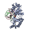
| Entry | Database: PDB / ID: 2oid | ||||||
|---|---|---|---|---|---|---|---|
| Title | Crystal structure of IRAK4 kinase domain complexed with AMPPNP | ||||||
 Components Components | Interleukin-1 receptor-associated kinase 4 | ||||||
 Keywords Keywords | TRANSFERASE / kinase | ||||||
| Function / homology |  Function and homology information Function and homology informationIRAK4 deficiency (TLR5) / MyD88 dependent cascade initiated on endosome / TRAF6 mediated induction of NFkB and MAP kinases upon TLR7/8 or 9 activation / MyD88 cascade initiated on plasma membrane / Toll signaling pathway / interleukin-33-mediated signaling pathway / neutrophil migration / toll-like receptor 9 signaling pathway / neutrophil mediated immunity / interleukin-1 receptor binding ...IRAK4 deficiency (TLR5) / MyD88 dependent cascade initiated on endosome / TRAF6 mediated induction of NFkB and MAP kinases upon TLR7/8 or 9 activation / MyD88 cascade initiated on plasma membrane / Toll signaling pathway / interleukin-33-mediated signaling pathway / neutrophil migration / toll-like receptor 9 signaling pathway / neutrophil mediated immunity / interleukin-1 receptor binding / interleukin-1-mediated signaling pathway / IRAK4 deficiency (TLR2/4) / MyD88:MAL(TIRAP) cascade initiated on plasma membrane / extrinsic component of plasma membrane / toll-like receptor 4 signaling pathway / MyD88-dependent toll-like receptor signaling pathway / toll-like receptor signaling pathway / JNK cascade / lipopolysaccharide-mediated signaling pathway / positive regulation of smooth muscle cell proliferation / TRAF6 mediated IRF7 activation in TLR7/8 or 9 signaling / cytokine-mediated signaling pathway / Interleukin-1 signaling / kinase activity / PIP3 activates AKT signaling / PI5P, PP2A and IER3 Regulate PI3K/AKT Signaling / positive regulation of canonical NF-kappaB signal transduction / non-specific serine/threonine protein kinase / endosome membrane / intracellular signal transduction / innate immune response / protein serine kinase activity / protein serine/threonine kinase activity / protein kinase binding / magnesium ion binding / cell surface / : / ATP binding / nucleus / plasma membrane / cytoplasm / cytosol Similarity search - Function | ||||||
| Biological species |  Homo sapiens (human) Homo sapiens (human) | ||||||
| Method |  X-RAY DIFFRACTION / SYNCHROTRON / DIFFERENCE FOURIER / Resolution: 2.3 Å X-RAY DIFFRACTION / SYNCHROTRON / DIFFERENCE FOURIER / Resolution: 2.3 Å | ||||||
 Authors Authors | Kuglstatter, A. / Villasenor, A.G. / Browner, M.F. | ||||||
 Citation Citation | Journal: J.Immunol. / Year: 2007 Title: Cutting Edge: IL-1 Receptor-Associated Kinase 4 Structures Reveal Novel Features and Multiple Conformations. Authors: Kuglstatter, A. / Villasenor, A.G. / Shaw, D. / Lee, S.W. / Tsing, S. / Niu, L. / Song, K.W. / Barnett, J.W. / Browner, M.F. | ||||||
| History |
|
- Structure visualization
| Structure viewer | Molecule: MolmilJmol/JSmol |
|---|
- Downloads & links
-Download
| PDBx/mmCIF format | 2oid.cif.gz | 230.5 KB | Display | PDBx/mmCIF format |
|---|---|---|---|---|
| PDB format | pdb2oid.ent.gz | 185.1 KB | Display | PDB format |
| PDBx/mmJSON format | 2oid.json.gz | Tree view | PDBx/mmJSON format | |
| Others |  Other downloads Other downloads |
-Validation report
| Arichive directory | https://data.pdbj.org/pub/pdb/validation_reports/oi/2oidftp://data.pdbj.org/pub/pdb/validation_reports/oi/2oid | HTTPS FTP |
|---|
-Related structure data
| Related structure data | 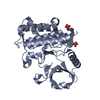 2oibSC 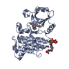 2oicC S: Starting model for refinement C: citing same article ( |
|---|---|
| Similar structure data |


-Links
 PDBj
PDBj


- Assembly
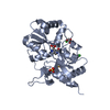
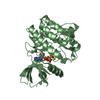
| Deposited unit | 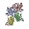
| ||||||||
|---|---|---|---|---|---|---|---|---|---|
| 1 | 
| ||||||||
| 2 | 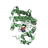
| ||||||||
| 3 | 
| ||||||||
| 4 | 
| ||||||||
| Unit cell |
| ||||||||
| Details | The asymmetric unit contains 4 kinase-ligand complexes, each of which represents one biological unit. |
-Components
| #1: Protein | Mass: 33956.195 Da / Num. of mol.: 4 / Fragment: kinase domain Source method: isolated from a genetically manipulated source Source: (gene. exp.) Homo sapiens (human) / Gene: IRAK4 / Production host:  unidentified baculovirus unidentified baculovirusReferences: UniProt: Q9NWZ3, non-specific serine/threonine protein kinase #2: Chemical | ChemComp-ANP /   Mass: 506.196 Da / Num. of mol.: 4 / Source method: obtained synthetically / Formula: C10H17N6O12P3 / Comment: AMP-PNP, energy-carrying molecule analogue*YM Mass: 506.196 Da / Num. of mol.: 4 / Source method: obtained synthetically / Formula: C10H17N6O12P3 / Comment: AMP-PNP, energy-carrying molecule analogue*YM#3: Water | ChemComp-HOH / |  Mass: 18.015 Da / Num. of mol.: 279 / Source method: isolated from a natural source / Formula: H2O Mass: 18.015 Da / Num. of mol.: 279 / Source method: isolated from a natural source / Formula: H2OHas protein modification | Y | |
|---|
-Experimental details
-Experiment
| Experiment | Method: X-RAY DIFFRACTION / Number of used crystals: 1 |
|---|
- Sample preparation
| Crystal | Density Matthews: 2.7 Å3/Da / Density % sol: 54.44 % |
|---|---|
| Crystal grow | Temperature: 293 K / Method: vapor diffusion, hanging drop / pH: 7 Details: 2.3M sodium malonate, 0.1M sodium acetate, 0.01M DTT, pH 7.0, VAPOR DIFFUSION, HANGING DROP, temperature 293K |
-Data collection
| Diffraction | Mean temperature: 200 K |
|---|---|
| Diffraction source | Source: SYNCHROTRON / Site: SSRL  / Beamline: BL9-1 / Wavelength: 0.97943 Å / Beamline: BL9-1 / Wavelength: 0.97943 Å |
| Detector | Type: ADSC QUANTUM 315 / Detector: CCD / Date: Jul 7, 2005 |
| Radiation | Protocol: SINGLE WAVELENGTH / Monochromatic (M) / Laue (L): M / Scattering type: x-ray |
| Radiation wavelength | Wavelength: 0.97943 Å / Relative weight: 1 |
| Reflection | Resolution: 2.3→50 Å / Num. obs: 63527 / % possible obs: 99.9 % / Observed criterion σ(F): 3 / Biso Wilson estimate: 50.8 Å2 / Rsym value: 0.112 / Net I/σ(I): 8.9 |
- Processing
| Software |
| ||||||||||||||||||||||||||||||||||||||||||||||||||||||||||||||||||||||||||||||||||||||||||
|---|---|---|---|---|---|---|---|---|---|---|---|---|---|---|---|---|---|---|---|---|---|---|---|---|---|---|---|---|---|---|---|---|---|---|---|---|---|---|---|---|---|---|---|---|---|---|---|---|---|---|---|---|---|---|---|---|---|---|---|---|---|---|---|---|---|---|---|---|---|---|---|---|---|---|---|---|---|---|---|---|---|---|---|---|---|---|---|---|---|---|---|
| Refinement | Method to determine structure: DIFFERENCE FOURIER Starting model: PDB ENTRY 2OIB Resolution: 2.3→49.94 Å / Cor.coef. Fo:Fc: 0.934 / Cor.coef. Fo:Fc free: 0.901 / SU B: 21.997 / SU ML: 0.238 / Cross valid method: THROUGHOUT / ESU R: 0.34 / ESU R Free: 0.266 / Stereochemistry target values: MAXIMUM LIKELIHOOD
| ||||||||||||||||||||||||||||||||||||||||||||||||||||||||||||||||||||||||||||||||||||||||||
| Solvent computation | Ion probe radii: 0.8 Å / Shrinkage radii: 0.8 Å / VDW probe radii: 1.2 Å / Solvent model: BABINET MODEL WITH MASK | ||||||||||||||||||||||||||||||||||||||||||||||||||||||||||||||||||||||||||||||||||||||||||
| Displacement parameters | Biso mean: 53.347 Å2
| ||||||||||||||||||||||||||||||||||||||||||||||||||||||||||||||||||||||||||||||||||||||||||
| Refinement step | Cycle: LAST / Resolution: 2.3→49.94 Å
| ||||||||||||||||||||||||||||||||||||||||||||||||||||||||||||||||||||||||||||||||||||||||||
| Refine LS restraints |
| ||||||||||||||||||||||||||||||||||||||||||||||||||||||||||||||||||||||||||||||||||||||||||
| LS refinement shell | Resolution: 2.3→2.36 Å / Total num. of bins used: 20
| ||||||||||||||||||||||||||||||||||||||||||||||||||||||||||||||||||||||||||||||||||||||||||
| Refinement TLS params. | Method: refined / Origin x: 22.1556 Å / Origin y: -16.4864 Å / Origin z: 17.7119 Å
| ||||||||||||||||||||||||||||||||||||||||||||||||||||||||||||||||||||||||||||||||||||||||||
| Refinement TLS group |
|
