 Movie
Movie Controller

[English] 日本語
 Yorodumi
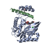
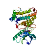
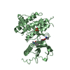
- PDB-4bgb: Nucleotide-bound closed form of a putative sugar kinase MK0840 fr... -
+ Open data

- Basic information
| Entry | Database: PDB / ID: 4bgb | ||||||
|---|---|---|---|---|---|---|---|
| Title | Nucleotide-bound closed form of a putative sugar kinase MK0840 from Methanopyrus kandleri | ||||||
 Components Components | PUTATIVE SUGAR KINASE MK0840 | ||||||
 Keywords Keywords | TRANSFERASE / PHOSPHOTRANSFER / PSEUDOMUREIN | ||||||
| Function / homology |  Function and homology information Function and homology informationamino sugar metabolic process / phosphotransferase activity, alcohol group as acceptor / peptidoglycan turnover / metallopeptidase activity / ATP binding / metal ion binding Similarity search - Function | ||||||
| Biological species |   METHANOPYRUS KANDLERI (archaea) METHANOPYRUS KANDLERI (archaea) | ||||||
| Method |  X-RAY DIFFRACTION / SYNCHROTRON / MOLECULAR REPLACEMENT / Resolution: 1.34 Å X-RAY DIFFRACTION / SYNCHROTRON / MOLECULAR REPLACEMENT / Resolution: 1.34 Å | ||||||
 Authors Authors | Schacherl, M. / Baumann, U. | ||||||
 Citation Citation | Journal: Acta Crystallogr.,Sect.D / Year: 2013 Title: Structural Characterization of the Ribonuclease H-Like Type Askha Superfamily Kinase Mk0840 from Methanopyrus Kandleri Authors: Schacherl, M. / Waltersperger, S.M. / Baumann, U. | ||||||
| History |
|
- Structure visualization
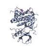
| Structure viewer | Molecule: MolmilJmol/JSmol |
|---|
- Downloads & links
-Download
| PDBx/mmCIF format | 4bgb.cif.gz | 284.5 KB | Display | PDBx/mmCIF format |
|---|---|---|---|---|
| PDB format | pdb4bgb.ent.gz | 227.7 KB | Display | PDB format |
| PDBx/mmJSON format | 4bgb.json.gz | Tree view | PDBx/mmJSON format | |
| Others |  Other downloads Other downloads |
-Validation report
| Arichive directory | https://data.pdbj.org/pub/pdb/validation_reports/bg/4bgbftp://data.pdbj.org/pub/pdb/validation_reports/bg/4bgb | HTTPS FTP |
|---|
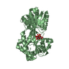
-Related structure data
| Related structure data | 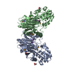 4bg8C 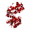 4bg9C 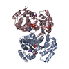 4bgaSC C: citing same article ( S: Starting model for refinement |
|---|---|
| Similar structure data |


-Links
 PDBj
PDBj- Assembly
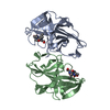
| Deposited unit | 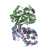
| ||||||||
|---|---|---|---|---|---|---|---|---|---|
| 1 | 
| ||||||||
| 2 | 
| ||||||||
| Unit cell |
|
-Components
-Protein , 1 types, 2 molecules AB
| #1: Protein | Mass: 35059.656 Da / Num. of mol.: 2 / Fragment: RESIDUES 37-358 Source method: isolated from a genetically manipulated source Source: (gene. exp.) METHANOPYRUS KANDLERI (archaea) / Strain: AV19 / Description: DSM6324 / Production host:  |
|---|
-Non-polymers , 6 types, 537 molecules 


| #2: Chemical |  Mass: 427.201 Da / Num. of mol.: 2 / Source method: obtained synthetically / Formula: C10H15N5O10P2 / Comment: ADP, energy-carrying molecule*YM Mass: 427.201 Da / Num. of mol.: 2 / Source method: obtained synthetically / Formula: C10H15N5O10P2 / Comment: ADP, energy-carrying molecule*YM#3: Chemical | ChemComp-GOL /  Mass: 92.094 Da / Num. of mol.: 7 / Source method: obtained synthetically / Formula: C3H8O3 Mass: 92.094 Da / Num. of mol.: 7 / Source method: obtained synthetically / Formula: C3H8O3#4: Chemical | ChemComp-K /  Mass: 39.098 Da / Num. of mol.: 4 / Source method: obtained synthetically / Formula: K Mass: 39.098 Da / Num. of mol.: 4 / Source method: obtained synthetically / Formula: K#5: Chemical | ChemComp-MG /  Mass: 24.305 Da / Num. of mol.: 4 / Source method: obtained synthetically / Formula: Mg Mass: 24.305 Da / Num. of mol.: 4 / Source method: obtained synthetically / Formula: Mg#6: Chemical | ChemComp-CA / |  Mass: 40.078 Da / Num. of mol.: 1 / Source method: obtained synthetically / Formula: Ca Mass: 40.078 Da / Num. of mol.: 1 / Source method: obtained synthetically / Formula: Ca#7: Water | ChemComp-HOH / | Mass: 18.015 Da / Num. of mol.: 519 / Source method: isolated from a natural source / Formula: H2O |
|---|
-Details
| Nonpolymer details | GLYCEROL (GOL): GOL7 ONLY PARTIALLY OCCUPIED |
|---|---|
| Sequence details | TRUNCATED CONSTRUCT LACKING THE FIRST 36 AMINO ACIDS |
-Experimental details
-Experiment
| Experiment | Method: X-RAY DIFFRACTION / Number of used crystals: 1 |
|---|
- Sample preparation
| Crystal | Density Matthews: 2.3 Å3/Da / Density % sol: 46.5 % Description: PARTIAL MODEL SOLVED BY MR. MODEL FOR SECOND PROTOMER - CLOSED FORM - BUILD BY WEB-BASED ARPWARP. MERGED DATASET FROM LOW-RESOLUTION AND HIGH-RESOLUTION PASS. |
|---|---|
| Crystal grow | pH: 4.6 Details: 0.14 M CACL2, 0.07 M SODIUM ACETATE PH 4.6, 10 % ISOPROPANOL AND 28% GLYCEROL |
-Data collection
| Diffraction | Mean temperature: 100 K |
|---|---|
| Diffraction source | Source: SYNCHROTRON / Site: SLS  / Beamline: X06DA / Wavelength: 1 / Beamline: X06DA / Wavelength: 1 |
| Detector | Type: MARMOSAIC 225 mm CCD / Detector: CCD / Date: Nov 8, 2011 |
| Radiation | Protocol: SINGLE WAVELENGTH / Monochromatic (M) / Laue (L): M / Scattering type: x-ray |
| Radiation wavelength | Wavelength: 1 Å / Relative weight: 1 |
| Reflection | Resolution: 1.34→45.2 Å / Num. obs: 154630 / % possible obs: 98.3 % / Observed criterion σ(I): 2 / Redundancy: 4.5 % / Biso Wilson estimate: 14.8 Å2 / Rmerge(I) obs: 0.04 / Net I/σ(I): 21.12 |
| Reflection shell | Resolution: 1.34→1.37 Å / Redundancy: 4 % / Rmerge(I) obs: 0.65 / Mean I/σ(I) obs: 2.61 / % possible all: 93.2 |
- Processing
| Software |
| |||||||||||||||||||||||||||||||||||||||||||||||||||||||||||||||||||||||||||||||||||||||||||||||||||||||||||||||||||||||||||||||||||||||||||||||||||||||||||||||||||||||||||||||||||||||||||||||||||||||||||||||||||||||||
|---|---|---|---|---|---|---|---|---|---|---|---|---|---|---|---|---|---|---|---|---|---|---|---|---|---|---|---|---|---|---|---|---|---|---|---|---|---|---|---|---|---|---|---|---|---|---|---|---|---|---|---|---|---|---|---|---|---|---|---|---|---|---|---|---|---|---|---|---|---|---|---|---|---|---|---|---|---|---|---|---|---|---|---|---|---|---|---|---|---|---|---|---|---|---|---|---|---|---|---|---|---|---|---|---|---|---|---|---|---|---|---|---|---|---|---|---|---|---|---|---|---|---|---|---|---|---|---|---|---|---|---|---|---|---|---|---|---|---|---|---|---|---|---|---|---|---|---|---|---|---|---|---|---|---|---|---|---|---|---|---|---|---|---|---|---|---|---|---|---|---|---|---|---|---|---|---|---|---|---|---|---|---|---|---|---|---|---|---|---|---|---|---|---|---|---|---|---|---|---|---|---|---|---|---|---|---|---|---|---|---|---|---|---|---|---|---|---|---|
| Refinement | Method to determine structure: MOLECULAR REPLACEMENT Starting model: PDB ENTRY 4BGA CHAIN A Resolution: 1.34→45.147 Å / SU ML: 0.13 / σ(F): 1.99 / Phase error: 20.02 / Stereochemistry target values: ML Details: FIRST 36 AMINO ACIDS OF THE PROTEIN ARE MISSING. GSH OVERHANG FROM THROMBIN-DIGESTED 6X-HIS TAG.
| |||||||||||||||||||||||||||||||||||||||||||||||||||||||||||||||||||||||||||||||||||||||||||||||||||||||||||||||||||||||||||||||||||||||||||||||||||||||||||||||||||||||||||||||||||||||||||||||||||||||||||||||||||||||||
| Solvent computation | Shrinkage radii: 1.1 Å / VDW probe radii: 1.3 Å / Solvent model: FLAT BULK SOLVENT MODEL / Bsol: 42.413 Å2 / ksol: 0.362 e/Å3 | |||||||||||||||||||||||||||||||||||||||||||||||||||||||||||||||||||||||||||||||||||||||||||||||||||||||||||||||||||||||||||||||||||||||||||||||||||||||||||||||||||||||||||||||||||||||||||||||||||||||||||||||||||||||||
| Displacement parameters | Biso mean: 27.09 Å2
| |||||||||||||||||||||||||||||||||||||||||||||||||||||||||||||||||||||||||||||||||||||||||||||||||||||||||||||||||||||||||||||||||||||||||||||||||||||||||||||||||||||||||||||||||||||||||||||||||||||||||||||||||||||||||
| Refinement step | Cycle: LAST / Resolution: 1.34→45.147 Å
| |||||||||||||||||||||||||||||||||||||||||||||||||||||||||||||||||||||||||||||||||||||||||||||||||||||||||||||||||||||||||||||||||||||||||||||||||||||||||||||||||||||||||||||||||||||||||||||||||||||||||||||||||||||||||
| Refine LS restraints |
| |||||||||||||||||||||||||||||||||||||||||||||||||||||||||||||||||||||||||||||||||||||||||||||||||||||||||||||||||||||||||||||||||||||||||||||||||||||||||||||||||||||||||||||||||||||||||||||||||||||||||||||||||||||||||
| LS refinement shell |
|
