 Movie
Movie Controller


+ Open data

- Basic information
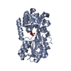
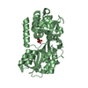
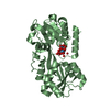
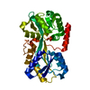
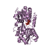
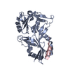
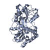
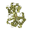
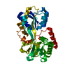
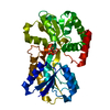
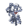
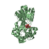
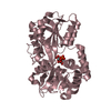
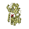
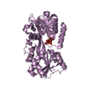
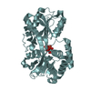
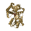
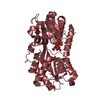
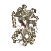
| Entry | Database: PDB / ID: 1xc1 | ||||||
|---|---|---|---|---|---|---|---|
| Title | Oxo Zirconium(IV) Cluster in the Ferric Binding Protein (FBP) | ||||||
 Components Components | periplasmic iron-binding protein | ||||||
 Keywords Keywords | METAL TRANSPORT / PERIPLASMIC FERRIC BINDING PROTEIN / ZIRCONIUM / METAL-OXO CLUSTER | ||||||
| Function / homology |  Function and homology information Function and homology informationiron ion transport / transmembrane transport / outer membrane-bounded periplasmic space / metal ion binding Similarity search - Function | ||||||
| Biological species |  Neisseria gonorrhoeae (bacteria) Neisseria gonorrhoeae (bacteria) | ||||||
| Method |  X-RAY DIFFRACTION / SYNCHROTRON / MOLECULAR REPLACEMENT / Resolution: 1.51 Å X-RAY DIFFRACTION / SYNCHROTRON / MOLECULAR REPLACEMENT / Resolution: 1.51 Å | ||||||
 Authors Authors | Zhong, W. / Alexeev, D. / Harvey, I. / Guo, M. / Hunter, D.J.B. / Zhu, H. / Campopiano, D.J. / Sadler, P.J. | ||||||
 Citation Citation | Journal: Angew.Chem.Int.Ed.Engl. / Year: 2004 Title: Assembly of an Oxo-Zirconium(IV) Cluster in a Protein Cleft Authors: Zhong, W. / Alexeev, D. / Harvey, I. / Guo, M. / Hunter, D.J.B. / Zhu, H. / Campopiano, D.J. / Sadler, P.J. #1: Journal: Biochem.J. / Year: 2003Title: Oxo-iron clusters in a bacterial iron-trafficking protein: new roles for a conserved motif Authors: Zhu, H. / Alexeev, D. / Hunter, D.J.B. / Campopiano, D.J. / Sadler, P.J. | ||||||
| History |
|
- Structure visualization
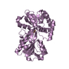
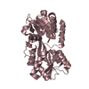
| Structure viewer | Molecule: MolmilJmol/JSmol |
|---|
- Downloads & links
-Download
| PDBx/mmCIF format | 1xc1.cif.gz | 546.4 KB | Display | PDBx/mmCIF format |
|---|---|---|---|---|
| PDB format | pdb1xc1.ent.gz | 449.3 KB | Display | PDB format |
| PDBx/mmJSON format | 1xc1.json.gz | Tree view | PDBx/mmJSON format | |
| Others |  Other downloads Other downloads |
-Validation report
| Arichive directory | https://data.pdbj.org/pub/pdb/validation_reports/xc/1xc1ftp://data.pdbj.org/pub/pdb/validation_reports/xc/1xc1 | HTTPS FTP |
|---|
-Related structure data
| Related structure data | 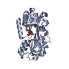 1o7tS S: Starting model for refinement |
|---|---|
| Similar structure data |


-Links
 PDBj
PDBj


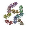
- Assembly
-Components
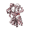
| #1: Protein | Mass: 33688.348 Da / Num. of mol.: 9 Source method: isolated from a genetically manipulated source Source: (gene. exp.) Neisseria gonorrhoeae (bacteria) / Gene: FBPA / Plasmid: PTRC99A-FBP-NG / Production host: #2: Chemical | ChemComp-ZRC /   Mass: 576.636 Da / Num. of mol.: 9 / Source method: obtained synthetically / Formula: O17PZr3 Mass: 576.636 Da / Num. of mol.: 9 / Source method: obtained synthetically / Formula: O17PZr3#3: Water | ChemComp-HOH / |  Mass: 18.015 Da / Num. of mol.: 974 / Source method: isolated from a natural source / Formula: H2O Mass: 18.015 Da / Num. of mol.: 974 / Source method: isolated from a natural source / Formula: H2O |
|---|
-Experimental details
-Experiment
| Experiment | Method: X-RAY DIFFRACTION / Number of used crystals: 1 |
|---|
- Sample preparation
| Crystal | Density Matthews: 2.32 Å3/Da / Density % sol: 46.48 % |
|---|---|
| Crystal grow | Temperature: 289 K / Method: vapor diffusion, hanging drop / pH: 8.1 Details: PEG 4000, NaCl, imidasole/malate, pH 8.1, VAPOR DIFFUSION, HANGING DROP, temperature 289K |
-Data collection
| Diffraction | Mean temperature: 100 K |
|---|---|
| Diffraction source | Source: SYNCHROTRON / Site: SRS  / Beamline: PX14.2 / Wavelength: 0.978 Å / Beamline: PX14.2 / Wavelength: 0.978 Å |
| Detector | Type: ADSC QUANTUM 4 / Detector: CCD / Date: Feb 3, 2002 / Details: mirrors |
| Radiation | Monochromator: SAGITALLY FOCUSED Si(111) / Protocol: SINGLE WAVELENGTH / Monochromatic (M) / Laue (L): M / Scattering type: x-ray |
| Radiation wavelength | Wavelength: 0.978 Å / Relative weight: 1 |
| Reflection | Resolution: 1.51→20 Å / Num. all: 426023 / Num. obs: 414535 / % possible obs: 98.5 % / Observed criterion σ(F): 0 / Observed criterion σ(I): 0 / Redundancy: 4.9 % / Biso Wilson estimate: 22.5 Å2 / Rmerge(I) obs: 0.095 / Rsym value: 0.095 / Net I/σ(I): 16.3 |
| Reflection shell | Resolution: 1.51→1.58 Å / Redundancy: 2.9 % / Rmerge(I) obs: 0.809 / Mean I/σ(I) obs: 1.3 / Num. unique all: 46715 / Rsym value: 0.81 / % possible all: 94.1 |
- Processing
| Software |
| |||||||||||||||||||||||||||||||||||||||||||||||||
|---|---|---|---|---|---|---|---|---|---|---|---|---|---|---|---|---|---|---|---|---|---|---|---|---|---|---|---|---|---|---|---|---|---|---|---|---|---|---|---|---|---|---|---|---|---|---|---|---|---|---|
| Refinement | Method to determine structure: MOLECULAR REPLACEMENT Starting model: PDB entry 1O7T Resolution: 1.51→20 Å / Isotropic thermal model: Isotropic / Cross valid method: THROUGHOUT / σ(F): 0 / σ(I): 0 / Stereochemistry target values: Engh & Huber Details: Twinned least squares refinement with the twinning fraction of 0.49
| |||||||||||||||||||||||||||||||||||||||||||||||||
| Displacement parameters | Biso mean: 26.8 Å2
| |||||||||||||||||||||||||||||||||||||||||||||||||
| Refinement step | Cycle: LAST / Resolution: 1.51→20 Å
| |||||||||||||||||||||||||||||||||||||||||||||||||
| Refine LS restraints |
| |||||||||||||||||||||||||||||||||||||||||||||||||
| LS refinement shell |
|
