 Movie
Movie Controller


+ Open data

- Basic information
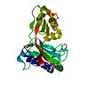
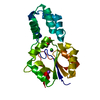
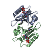
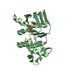
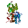
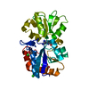
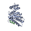
| Entry | Database: PDB / ID: 1a7x | ||||||
|---|---|---|---|---|---|---|---|
| Title | FKBP12-FK1012 COMPLEX | ||||||
 Components Components | FKBP12 | ||||||
 Keywords Keywords | ISOMERASE / IMMUNOPHILIN | ||||||
| Function / homology |  Function and homology information Function and homology informationmacrolide binding / activin receptor binding / regulation of skeletal muscle contraction by regulation of release of sequestered calcium ion / cytoplasmic side of membrane / transforming growth factor beta receptor binding / TGFBR1 LBD Mutants in Cancer / type I transforming growth factor beta receptor binding / negative regulation of activin receptor signaling pathway / signaling receptor inhibitor activity / heart trabecula formation ...macrolide binding / activin receptor binding / regulation of skeletal muscle contraction by regulation of release of sequestered calcium ion / cytoplasmic side of membrane / transforming growth factor beta receptor binding / TGFBR1 LBD Mutants in Cancer / type I transforming growth factor beta receptor binding / negative regulation of activin receptor signaling pathway / signaling receptor inhibitor activity / heart trabecula formation / I-SMAD binding / regulation of amyloid precursor protein catabolic process / terminal cisterna / ryanodine receptor complex / 'de novo' protein folding / ventricular cardiac muscle tissue morphogenesis / FK506 binding / TGF-beta receptor signaling activates SMADs / mTORC1-mediated signalling / Calcineurin activates NFAT / regulation of immune response / heart morphogenesis / supramolecular fiber organization / regulation of cardiac muscle contraction by regulation of the release of sequestered calcium ion / sarcoplasmic reticulum membrane / T cell activation / peptidylprolyl isomerase / sarcoplasmic reticulum / peptidyl-prolyl cis-trans isomerase activity / TGF-beta receptor signaling in EMT (epithelial to mesenchymal transition) / calcium channel regulator activity / negative regulation of transforming growth factor beta receptor signaling pathway / protein maturation / Z disc / SARS-CoV-1 activates/modulates innate immune responses / regulation of protein localization / protein refolding / protein folding / amyloid fibril formation / Potential therapeutics for SARS / transmembrane transporter binding / positive regulation of canonical NF-kappaB signal transduction / membrane / cytoplasm / cytosol Similarity search - Function | ||||||
| Biological species |  Homo sapiens (human) Homo sapiens (human) | ||||||
| Method |  X-RAY DIFFRACTION / MOLECULAR REPLACEMENT / Resolution: 2 Å X-RAY DIFFRACTION / MOLECULAR REPLACEMENT / Resolution: 2 Å | ||||||
 Authors Authors | Schultz, L.W. / Clardy, J. | ||||||
 Citation Citation | Journal: Bioorg.Med.Chem.Lett. / Year: 1998 Title: Chemical inducers of dimerization: the atomic structure of FKBP12-FK1012A-FKBP12. Authors: Schultz, L.W. / Clardy, J. #1: Journal: Science / Year: 1991Title: Atomic Structure of Fkbp-Fk506, an Immunophilin-Immunosuppressant Complex Authors: Van Duyne, G.D. / Standaert, R.F. / Karplus, P.A. / Schreiber, S.L. / Clardy, J. #2: Journal: Nature / Year: 1990Title: Molecular Cloning and Overexpression of the Human Fk506-Binding Protein Fkbp Authors: Standaert, R.F. / Galat, A. / Verdine, G.L. / Schreiber, S.L. | ||||||
| History |
|
- Structure visualization
| Structure viewer | Molecule: MolmilJmol/JSmol |
|---|
- Downloads & links
-Download
| PDBx/mmCIF format | 1a7x.cif.gz | 68.3 KB | Display | PDBx/mmCIF format |
|---|---|---|---|---|
| PDB format | pdb1a7x.ent.gz | 50.5 KB | Display | PDB format |
| PDBx/mmJSON format | 1a7x.json.gz | Tree view | PDBx/mmJSON format | |
| Others |  Other downloads Other downloads |
-Validation report
| Arichive directory | https://data.pdbj.org/pub/pdb/validation_reports/a7/1a7xftp://data.pdbj.org/pub/pdb/validation_reports/a7/1a7x | HTTPS FTP |
|---|
-Related structure data
| Related structure data |  1fkfS S: Starting model for refinement |
|---|---|
| Similar structure data |


-Links
 PDBj
PDBj


- Assembly
| Deposited unit | 
| ||||||||
|---|---|---|---|---|---|---|---|---|---|
| 1 |
| ||||||||
| Unit cell |
| ||||||||
| Components on special symmetry positions |
|
-Components
| #1: Protein | Mass: 11836.508 Da / Num. of mol.: 2 Source method: isolated from a genetically manipulated source Source: (gene. exp.) Homo sapiens (human) / Cellular location: CYTOPLASM / Production host:  #2: Chemical | ChemComp-FKA / |   Mass: 941.154 Da / Num. of mol.: 1 / Source method: obtained synthetically / Formula: C51H76N2O14 Mass: 941.154 Da / Num. of mol.: 1 / Source method: obtained synthetically / Formula: C51H76N2O14#3: Water | ChemComp-HOH / |  Mass: 18.015 Da / Num. of mol.: 177 / Source method: isolated from a natural source / Formula: H2O Mass: 18.015 Da / Num. of mol.: 177 / Source method: isolated from a natural source / Formula: H2O |
|---|
-Experimental details
-Experiment
| Experiment | Method: X-RAY DIFFRACTION / Number of used crystals: 1 |
|---|
- Sample preparation
| Crystal | Density Matthews: 2.66 Å3/Da / Density % sol: 50 % | |||||||||||||||||||||||||
|---|---|---|---|---|---|---|---|---|---|---|---|---|---|---|---|---|---|---|---|---|---|---|---|---|---|---|
| Crystal grow | Method: vapor diffusion, hanging drop / pH: 4.6 Details: A 10MG/ML SOLUTION OF FK1012A IN MEOH WAS ADDED IN A 1:2 MOLAR RATIO TO A 10MG/ML SOLUTION OF FKBP12 IN 10MM TRIS PH 8.2. THE SAMPLE WAS GENTLY MIXED AND ALLOWED TO INCUBATE OVERNIGHT TO ...Details: A 10MG/ML SOLUTION OF FK1012A IN MEOH WAS ADDED IN A 1:2 MOLAR RATIO TO A 10MG/ML SOLUTION OF FKBP12 IN 10MM TRIS PH 8.2. THE SAMPLE WAS GENTLY MIXED AND ALLOWED TO INCUBATE OVERNIGHT TO ENSURE COMPLETE BINDING. CRYSTALS WERE GROWN USING THE HANGING DROP METHOD WITH 0.5ML RESERVOIR CONSISTING OF 5.1M SODIUM FORMATE AND 0.1M SODIUM ACETATE PH 4.6. THE DROPS CONSISTED OF 3UL OF PROTEIN AND 3UL OF RESERVOIR SOLUTION., vapor diffusion - hanging drop PH range: 5.1-8.2 | |||||||||||||||||||||||||
| Crystal grow | *PLUS Method: vapor diffusion, hanging dropDetails: drop contains equal volume of the reservoir solution | |||||||||||||||||||||||||
| Components of the solutions | *PLUS
|
-Data collection
| Diffraction | Mean temperature: 294 K |
|---|---|
| Diffraction source | Source: ROTATING ANODE / Type: RIGAKU RUH2R / Wavelength: 1.5418 |
| Detector | Type: XUONG-HAMLIN MULTIWIRE / Detector: AREA DETECTOR / Date: Apr 10, 1995 |
| Radiation | Monochromator: GRAPHITE(002) / Monochromatic (M) / Laue (L): M / Scattering type: x-ray |
| Radiation wavelength | Wavelength: 1.5418 Å / Relative weight: 1 |
| Reflection | Resolution: 2→20 Å / Num. obs: 15896 / % possible obs: 91 % / Observed criterion σ(I): 2 / Redundancy: 2 % / Rsym value: 0.046 / Net I/σ(I): 14 |
| Reflection | *PLUS Rmerge(I) obs: 0.046 |
- Processing
| Software |
| ||||||||||||||||||||||||||||||||||||||||||||||||||||||||||||
|---|---|---|---|---|---|---|---|---|---|---|---|---|---|---|---|---|---|---|---|---|---|---|---|---|---|---|---|---|---|---|---|---|---|---|---|---|---|---|---|---|---|---|---|---|---|---|---|---|---|---|---|---|---|---|---|---|---|---|---|---|---|
| Refinement | Method to determine structure: MOLECULAR REPLACEMENT Starting model: PDB ENTRY 1FKF Resolution: 2→8 Å / σ(F): 2 | ||||||||||||||||||||||||||||||||||||||||||||||||||||||||||||
| Refinement step | Cycle: LAST / Resolution: 2→8 Å
| ||||||||||||||||||||||||||||||||||||||||||||||||||||||||||||
| Refine LS restraints |
| ||||||||||||||||||||||||||||||||||||||||||||||||||||||||||||
| Xplor file |
| ||||||||||||||||||||||||||||||||||||||||||||||||||||||||||||
| Software | *PLUS Name: X-PLOR / Version: 3 / Classification: refinement | ||||||||||||||||||||||||||||||||||||||||||||||||||||||||||||
| Refinement | *PLUS Rfactor obs: 0.173 | ||||||||||||||||||||||||||||||||||||||||||||||||||||||||||||
| Solvent computation | *PLUS | ||||||||||||||||||||||||||||||||||||||||||||||||||||||||||||
| Displacement parameters | *PLUS |
