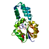
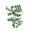
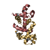
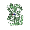
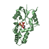
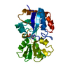
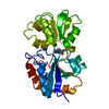
Entry Database : PDB / ID : 4j8sTitle Crystal structure of human CNOT1 MIF4G domain in complex with a TTP peptide CCR4-NOT transcription complex subunit 1 Tristetraprolin Keywords / / / / Function / homology Function Domain/homology Component
/ / / / / / / / / / / / / / / / / / / / / / / / / / / / / / / / / / / / / / / / / / / / / / / / / / / / / / / / / / / / / / / / / / / / / / / / / / / / / / / / / / / / / / / / / / / / / / / / / / / / / / / / / / / / / / / / / / / / / / / / / / / / Biological species Homo sapiens (human)HOMO SAPIENS (human)Method / / Resolution : 1.55 Å Authors Frank, F. / Fabian, M.R. / Rouya, C. / Siddiqui, N. / Lai, W.S. / Karetnikov, A. / Blackshear, P.J. / Sonenberg, N. / Nagar, B. Journal : Nat.Struct.Mol.Biol. / Year : 2013Title : Structural basis for the recruitment of the human CCR4-NOT deadenylase complex by tristetraprolin.Authors : Fabian, M.R. / Frank, F. / Rouya, C. / Siddiqui, N. / Lai, W.S. / Karetnikov, A. / Blackshear, P.J. / Nagar, B. / Sonenberg, N. History Deposition Feb 14, 2013 Deposition site / Processing site Revision 1.0 May 8, 2013 Provider / Type Revision 1.1 Jul 24, 2013 Group Revision 1.2 May 25, 2016 Group Revision 1.3 Feb 28, 2024 Group / Database referencesCategory chem_comp_atom / chem_comp_bond ... chem_comp_atom / chem_comp_bond / database_2 / struct_ref_seq_dif Item / _database_2.pdbx_database_accession / _struct_ref_seq_dif.details
Show all Show less
 Movie
Movie Controller

 Yorodumi
 Open data
 Basic information
 Components
 Keywords
 Function and homology information
 Homo sapiens (human)
 X-RAY DIFFRACTION / Sulfur SAD / Resolution: 1.55 Å
  Authors
 Citation
 Structure visualization
 Downloads & links
 Other downloads
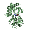


 PDBj
PDBj


 Assembly
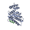

 Mass: 18.015 Da / Num. of mol.: 237 / Source method: isolated from a natural source / Formula: H2O
 Sample preparation
 Processing