 Movie
Movie Controller

[English] 日本語
 Yorodumi
- PDB-1wnh: Crystal structure of mouse Latexin (tissue carboxypeptidase inhibitor) -
+ Open data

- Basic information
| Entry | Database: PDB / ID: 1wnh | ||||||
|---|---|---|---|---|---|---|---|
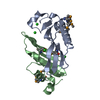
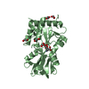
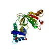
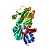
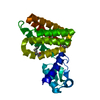
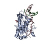
| Title | Crystal structure of mouse Latexin (tissue carboxypeptidase inhibitor) | ||||||
 Components Components | Latexin | ||||||
 Keywords Keywords | HYDROLASE INHIBITOR / bi-cystatin fold / cis-proline | ||||||
| Function / homology |  Function and homology information Function and homology informationmetalloendopeptidase inhibitor activity / detection of temperature stimulus involved in sensory perception of pain / heparin binding / inflammatory response / : / cytoplasm Similarity search - Function | ||||||
| Biological species |  | ||||||
| Method |  X-RAY DIFFRACTION / SYNCHROTRON / MAD / Resolution: 1.83 Å X-RAY DIFFRACTION / SYNCHROTRON / MAD / Resolution: 1.83 Å | ||||||
 Authors Authors | Aagaard, A. / Listwan, P. / Cowieson, N. / Huber, T. / Ravasi, T. / Wells, C.A. / Flanagan, J.U. / Hume, D.A. / Kobe, B. / Martin, J.L. | ||||||
 Citation Citation | Journal: Structure / Year: 2005 Title: An Inflammatory Role for the Mammalian Carboxypeptidase Inhibitor Latexin: Relationship to Cystatins and the Tumor Suppressor TIG1 Authors: Aagaard, A. / Listwan, P. / Cowieson, N. / Huber, T. / Ravasi, T. / Wells, C.A. / Flanagan, J.U. / Kellie, S. / Hume, D.A. / Kobe, B. / Martin, J.L. | ||||||
| History |
|
- Structure visualization
| Structure viewer | Molecule: MolmilJmol/JSmol |
|---|
- Downloads & links
-Download
| PDBx/mmCIF format | 1wnh.cif.gz | 62.6 KB | Display | PDBx/mmCIF format |
|---|---|---|---|---|
| PDB format | pdb1wnh.ent.gz | 46.4 KB | Display | PDB format |
| PDBx/mmJSON format | 1wnh.json.gz | Tree view | PDBx/mmJSON format | |
| Others |  Other downloads Other downloads |
-Validation report
| Arichive directory | https://data.pdbj.org/pub/pdb/validation_reports/wn/1wnhftp://data.pdbj.org/pub/pdb/validation_reports/wn/1wnh | HTTPS FTP |
|---|
-Related structure data
| Similar structure data |
|---|


-Links
 PDBj
PDBj- Assembly
| Deposited unit | 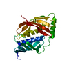
| ||||||||
|---|---|---|---|---|---|---|---|---|---|
| 1 |
| ||||||||
| Unit cell |
| ||||||||
| Details | The molecule in the asymmetric unit is assumed to represent the biological unit |
-Components
| #1: Protein | Mass: 25838.109 Da / Num. of mol.: 1 Source method: isolated from a genetically manipulated source Source: (gene. exp.)  |
|---|---|
| #2: Water | ChemComp-HOH /  Mass: 18.015 Da / Num. of mol.: 184 / Source method: isolated from a natural source / Formula: H2O Mass: 18.015 Da / Num. of mol.: 184 / Source method: isolated from a natural source / Formula: H2O |
-Experimental details
-Experiment
| Experiment | Method: X-RAY DIFFRACTION / Number of used crystals: 1 |
|---|
- Sample preparation
| Crystal | Density Matthews: 2.9 Å3/Da / Density % sol: 58 % |
|---|---|
| Crystal grow | Temperature: 293 K / Method: vapor diffusion, hanging drop / pH: 6.5 Details: 1.8M ammonium sulfate, 0.1M cacodylate, pH 6.5, VAPOR DIFFUSION, HANGING DROP, temperature 293K |
-Data collection
| Diffraction | Mean temperature: 100 K | |||||||||||||||
|---|---|---|---|---|---|---|---|---|---|---|---|---|---|---|---|---|
| Diffraction source | Source: SYNCHROTRON / Site: ALS  / Beamline: 8.2.2 / Wavelength: 1.0781, 0.9794, 0.9796, 0.9077 / Beamline: 8.2.2 / Wavelength: 1.0781, 0.9794, 0.9796, 0.9077 | |||||||||||||||
| Detector | Type: ADSC QUANTUM 4 / Detector: CCD / Date: Mar 8, 2003 | |||||||||||||||
| Radiation | Monochromator: Double-crystal, Si(111) / Protocol: MAD / Monochromatic (M) / Laue (L): M / Scattering type: x-ray | |||||||||||||||
| Radiation wavelength |
| |||||||||||||||
| Reflection | Resolution: 1.83→50 Å / Num. obs: 29633 / % possible obs: 99.7 % / Redundancy: 14 % / Biso Wilson estimate: 27.4 Å2 / Rmerge(I) obs: 0.039 / Net I/σ(I): 19.9 | |||||||||||||||
| Reflection shell | Resolution: 1.83→1.9 Å / Rmerge(I) obs: 0.422 / Mean I/σ(I) obs: 4.5 / % possible all: 100 |
- Processing
| Software |
| ||||||||||||||||||||||||||||||||||||
|---|---|---|---|---|---|---|---|---|---|---|---|---|---|---|---|---|---|---|---|---|---|---|---|---|---|---|---|---|---|---|---|---|---|---|---|---|---|
| Refinement | Method to determine structure: MAD / Resolution: 1.83→46.47 Å / Rfactor Rfree error: 0.004 / Data cutoff high absF: 606163.56 / Data cutoff low absF: 0 / Isotropic thermal model: RESTRAINED / Cross valid method: THROUGHOUT / σ(F): 0 / Stereochemistry target values: Engh & Huber
| ||||||||||||||||||||||||||||||||||||
| Solvent computation | Solvent model: FLAT MODEL / Bsol: 69.986 Å2 / ksol: 0.370558 e/Å3 | ||||||||||||||||||||||||||||||||||||
| Displacement parameters | Biso mean: 39.9 Å2
| ||||||||||||||||||||||||||||||||||||
| Refine analyze |
| ||||||||||||||||||||||||||||||||||||
| Refinement step | Cycle: LAST / Resolution: 1.83→46.47 Å
| ||||||||||||||||||||||||||||||||||||
| Refine LS restraints |
| ||||||||||||||||||||||||||||||||||||
| LS refinement shell | Resolution: 1.83→1.94 Å / Rfactor Rfree error: 0.013 / Total num. of bins used: 6
| ||||||||||||||||||||||||||||||||||||
| Xplor file |
|
