Knl1/Spc105 complex / positive regulation of meiosis I spindle assembly checkpoint / homologous chromosome orientation in meiotic metaphase I / mitotic checkpoint complex / acrosome assembly / regulation of mitotic cell cycle spindle assembly checkpoint / meiotic sister chromatid cohesion, centromeric / Inactivation of APC/C via direct inhibition of the APC/C complex / APC/C:Cdc20 mediated degradation of mitotic proteins / attachment of spindle microtubules to kinetochore ...Knl1/Spc105 complex / positive regulation of meiosis I spindle assembly checkpoint / homologous chromosome orientation in meiotic metaphase I / mitotic checkpoint complex / acrosome assembly / regulation of mitotic cell cycle spindle assembly checkpoint / meiotic sister chromatid cohesion, centromeric / Inactivation of APC/C via direct inhibition of the APC/C complex / APC/C:Cdc20 mediated degradation of mitotic proteins / attachment of spindle microtubules to kinetochore / anaphase-promoting complex / metaphase/anaphase transition of mitotic cell cycle / protein localization to chromosome, centromeric region / outer kinetochore / protein localization to kinetochore / mitotic spindle assembly checkpoint signaling / mitotic sister chromatid segregation / Amplification of signal from unattached kinetochores via a MAD2 inhibitory signal / Mitotic Prometaphase / Deposition of new CENPA-containing nucleosomes at the centromere / EML4 and NUDC in mitotic spindle formation / acrosomal vesicle / APC-Cdc20 mediated degradation of Nek2A / Resolution of Sister Chromatid Cohesion / Cdc20:Phospho-APC/C mediated degradation of Cyclin A / RHO GTPases Activate Formins / kinetochore / spindle / Separation of Sister Chromatids / microtubule binding / protein kinase activity / non-specific serine/threonine protein kinase / nuclear body / ciliary basal body / protein serine kinase activity / cell division / protein serine/threonine kinase activity / apoptotic process / centrosome / perinuclear region of cytoplasm / nucleoplasm / ATP binding / nucleus / cytoplasm / cytosol Similarity search - Function
Mitoticcheckpointserine/threonine-proteinkinaseBUB1beta / MAD3/BUB1-related protein kinase / hBUBR1 / Mitotic checkpoint kinase MAD3L / Protein SSK1
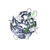
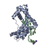
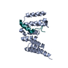
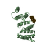
Mass: 20578.621 Da / Num. of mol.: 2 / Fragment: UNP residues 67-220 Source method: isolated from a genetically manipulated source Source: (gene. exp.) Homo sapiens (human) / Gene: BUB1B, BUBR1, MAD3L, SSK1 / Plasmid: PGEX / Production host: Escherichia coli (E. coli) / Strain (production host): BL21 References: UniProt: O60566, non-specific serine/threonine protein kinase
#2: Protein/peptide
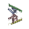
ProteinCASC5 / ALL1-fused gene from chromosome 15q14 protein / AF15q14 / Bub-linking kinetochore protein / Blinkin ...ALL1-fused gene from chromosome 15q14 protein / AF15q14 / Bub-linking kinetochore protein / Blinkin / Cancer susceptibility candidate gene 5 protein / Cancer/testis antigen 29 / CT29 / Kinetochore-null protein 1 / Protein D40/AF15q14
Mass: 2657.010 Da / Num. of mol.: 2 / Fragment: UNP residues 234-252 Source method: isolated from a genetically manipulated source Source: (gene. exp.) Homo sapiens (human) / Gene: CASC5, KIAA1570, KNL1 / Plasmid: PGEX / Production host: Escherichia coli (E. coli) / Strain (production host): BL21 / References: UniProt: Q8NG31
Resolution: 2.2→38.2 Å / Cor.coef. Fo:Fc: 0.937 / Cor.coef. Fo:Fc free: 0.902 / SU B: 13.612 / SU ML: 0.159 / Cross valid method: THROUGHOUT / ESU R Free: 0.225 / Stereochemistry target values: MAXIMUM LIKELIHOOD / Details: HYDROGENS HAVE BEEN ADDED IN THE RIDING POSITIONS
Rfactor
Num. reflection
% reflection
Selection details
Rfree
0.24665
985
5.1 %
RANDOM
Rwork
0.19343
-
-
-
all
0.19626
18292
-
-
obs
0.19626
18278
99.92 %
-
Solvent computation
Ion probe radii: 0.8 Å / Shrinkage radii: 0.8 Å / VDW probe radii: 1.4 Å / Solvent model: MASK
Displacement parameters
Biso mean: 24.773 Å2
Baniso -1
Baniso -2
Baniso -3
1-
-0.02 Å2
-0 Å2
0 Å2
2-
-
0.01 Å2
0 Å2
3-
-
-
0.01 Å2
Refinement step
Cycle: LAST / Resolution: 2.2→38.2 Å
Protein
Nucleic acid
Ligand
Solvent
Total
Num. atoms
2756
0
0
250
3006
Refine LS restraints
Refine-ID
Type
Dev ideal
Dev ideal target
Number
X-RAY DIFFRACTION
r_bond_refined_d
0.011
0.022
2866
X-RAY DIFFRACTION
r_angle_refined_deg
1.116
1.93
3875
X-RAY DIFFRACTION
r_dihedral_angle_1_deg
5.113
5
340
X-RAY DIFFRACTION
r_dihedral_angle_2_deg
40.501
23.908
174
X-RAY DIFFRACTION
r_dihedral_angle_3_deg
15.141
15
491
X-RAY DIFFRACTION
r_dihedral_angle_4_deg
17.699
15
28
X-RAY DIFFRACTION
r_chiral_restr
0.074
0.2
385
X-RAY DIFFRACTION
r_gen_planes_refined
0.005
0.021
2290
X-RAY DIFFRACTION
r_mcbond_it
0.603
1.5
1667
X-RAY DIFFRACTION
r_mcangle_it
1.133
2
2658
X-RAY DIFFRACTION
r_scbond_it
1.655
3
1199
X-RAY DIFFRACTION
r_scangle_it
2.733
4.5
1211
LS refinement shell
Resolution: 2.2→2.257 Å / Total num. of bins used: 20
Rfactor
Num. reflection
% reflection
Rfree
0.28
70
-
Rwork
0.2
1347
-
obs
-
-
100 %
Refinement TLS params.
Method: refined / Refine-ID: X-RAY DIFFRACTION
ID
L11 (°2)
L12 (°2)
L13 (°2)
L22 (°2)
L23 (°2)
L33 (°2)
S11 (Å °)
S12 (Å °)
S13 (Å °)
S21 (Å °)
S22 (Å °)
S23 (Å °)
S31 (Å °)
S32 (Å °)
S33 (Å °)
T11 (Å2)
T12 (Å2)
T13 (Å2)
T22 (Å2)
T23 (Å2)
T33 (Å2)
Origin x (Å)
Origin y (Å)
Origin z (Å)
1
0.3243
0.135
0.4737
0.1335
0.1689
0.6627
0.0106
-0.0138
0.005
0.0193
-0.0257
0.0016
0.0243
-0.0326
0.0152
0.1553
-0.0096
-0.0058
0.1385
0.0003
0.1604
14.7898
-0.1732
31.4405
2
0.3099
0.2216
-0.5514
0.1606
-0.4061
1.4972
0.0035
-0.0092
-0.0182
-0.0026
-0.029
0.0019
-0.0591
-0.0215
0.0255
0.16
-0.0028
-0.0101
0.1301
-0.0043
0.1499
47.6858
9.7792
13.6067
3
10.8371
-5.6074
-0.2818
9.1263
1.7082
0.0347
0.2274
0.3935
-0.1987
-0.6638
-0.1728
0.1254
-0.1653
0.139
-0.0546
0.1951
-0.033
0.003
0.2051
-0.0424
0.1001
19.09
7.7074
17.5814
4
17.5119
0.8577
-1.9466
23.3269
15.4918
10.9089
0.4008
-0.0045
-0.1507
-0.6329
0.0663
0.2502
-0.2076
-1.3103
-0.4671
0.0294
-0.208
-0.1167
0.8969
0.4503
0.1625
34.223
5.7306
6.0214
Refinement TLS group
ID
Refine-ID
Refine TLS-ID
Auth asym-ID
Auth seq-ID
1
X-RAY DIFFRACTION
1
A
54 - 205
2
X-RAY DIFFRACTION
2
B
55 - 203
3
X-RAY DIFFRACTION
3
X
2 - 19
4
X-RAY DIFFRACTION
4
Y
6 - 18
+
About Yorodumi
-
News
-
Feb 9, 2022. New format data for meta-information of EMDB entries
New format data for meta-information of EMDB entries
Version 3 of the EMDB header file is now the official format.
The previous official version 1.9 will be removed from the archive.
In the structure databanks used in Yorodumi, some data are registered as the other names, "COVID-19 virus" and "2019-nCoV". Here are the details of the virus and the list of structure data.
Jan 31, 2019. EMDB accession codes are about to change! (news from PDBe EMDB page)
EMDB accession codes are about to change! (news from PDBe EMDB page)
The allocation of 4 digits for EMDB accession codes will soon come to an end. Whilst these codes will remain in use, new EMDB accession codes will include an additional digit and will expand incrementally as the available range of codes is exhausted. The current 4-digit format prefixed with “EMD-” (i.e. EMD-XXXX) will advance to a 5-digit format (i.e. EMD-XXXXX), and so on. It is currently estimated that the 4-digit codes will be depleted around Spring 2019, at which point the 5-digit format will come into force.
The EM Navigator/Yorodumi systems omit the EMD- prefix.
Related info.:Q: What is EMD? / ID/Accession-code notation in Yorodumi/EM Navigator
Yorodumi is a browser for structure data from EMDB, PDB, SASBDB, etc.
This page is also the successor to EM Navigator detail page, and also detail information page/front-end page for Omokage search.
The word "yorodu" (or yorozu) is an old Japanese word meaning "ten thousand". "mi" (miru) is to see.
Related info.:EMDB / PDB / SASBDB / Comparison of 3 databanks / Yorodumi Search / Aug 31, 2016. New EM Navigator & Yorodumi / Yorodumi Papers / Jmol/JSmol / Function and homology information / Changes in new EM Navigator and Yorodumi
 Movie
Movie Controller


 Open data
 Basic information
 Components
 Keywords
 Function and homology information
 Homo sapiens (human)
 X-RAY DIFFRACTION /
  Authors
 Citation
 Structure visualization
 Downloads & links
 Other downloads


 PDBj
PDBj


 Assembly

 Mass: 18.015 Da / Num. of mol.: 250 / Source method: isolated from a natural source / Formula: H2O
 Sample preparation
 Processing