 Movie
Movie Controller

[English] 日本語
 Yorodumi
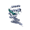
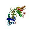
- PDB-1eaj: DIMERIC STRUCTURE OF THE COXSACKIE VIRUS AND ADENOVIRUS RECEPTOR ... -
+ Open data

- Basic information
| Entry | Database: PDB / ID: 1eaj | ||||||
|---|---|---|---|---|---|---|---|
| Title | DIMERIC STRUCTURE OF THE COXSACKIE VIRUS AND ADENOVIRUS RECEPTOR D1 DOMAIN AT 1.35 ANGSTROM RESOLUTION | ||||||
 Components Components | COXSACKIE VIRUS AND ADENOVIRUS RECEPTOR | ||||||
 Keywords Keywords | VIRUS/VIRAL PROTEIN RECEPTOR / IMMUNOGLOBULIN V DOMAIN FOLD / SYMMETRIC DIMER / VIRUS-VIRAL PROTEIN RECEPTOR complex | ||||||
| Function / homology |  Function and homology information Function and homology informationAV node cell-bundle of His cell adhesion involved in cell communication / cell adhesive protein binding involved in AV node cell-bundle of His cell communication / AV node cell to bundle of His cell communication / homotypic cell-cell adhesion / epithelial structure maintenance / regulation of AV node cell action potential / gamma-delta T cell activation / apicolateral plasma membrane / primordial germ cell migration / connexin binding ...AV node cell-bundle of His cell adhesion involved in cell communication / cell adhesive protein binding involved in AV node cell-bundle of His cell communication / AV node cell to bundle of His cell communication / homotypic cell-cell adhesion / epithelial structure maintenance / regulation of AV node cell action potential / gamma-delta T cell activation / apicolateral plasma membrane / primordial germ cell migration / connexin binding / cell-cell junction organization / transepithelial transport / heterophilic cell-cell adhesion / cardiac muscle cell development / intercalated disc / bicellular tight junction / neutrophil chemotaxis / cell adhesion molecule binding / acrosomal vesicle / Cell surface interactions at the vascular wall / filopodium / adherens junction / PDZ domain binding / neuromuscular junction / mitochondrion organization / beta-catenin binding / integrin binding / Immunoregulatory interactions between a Lymphoid and a non-Lymphoid cell / cell-cell junction / cell junction / heart development / virus receptor activity / growth cone / actin cytoskeleton organization / cell body / defense response to virus / basolateral plasma membrane / neuron projection / membrane raft / signaling receptor binding / protein-containing complex / : / extracellular region / nucleoplasm / identical protein binding / plasma membrane / cytoplasm Similarity search - Function | ||||||
| Biological species |  HOMO SAPIENS (human) HOMO SAPIENS (human) | ||||||
| Method |  X-RAY DIFFRACTION / SYNCHROTRON / MOLECULAR REPLACEMENT / Resolution: 1.35 Å X-RAY DIFFRACTION / SYNCHROTRON / MOLECULAR REPLACEMENT / Resolution: 1.35 Å | ||||||
 Authors Authors | van Raaij, M.J. / Cusack, S. | ||||||
 Citation Citation | Journal: Structure / Year: 2000 Title: Dimeric structure of the coxsackievirus and adenovirus receptor D1 domain at 1.7 A resolution. Authors: van Raaij, M.J. / Chouin, E. / van der Zandt, H. / Bergelson, J.M. / Cusack, S. | ||||||
| History |
|
- Structure visualization
| Structure viewer | Molecule: MolmilJmol/JSmol |
|---|
- Downloads & links
-Download
| PDBx/mmCIF format | 1eaj.cif.gz | 123.1 KB | Display | PDBx/mmCIF format |
|---|---|---|---|---|
| PDB format | pdb1eaj.ent.gz | 96.8 KB | Display | PDB format |
| PDBx/mmJSON format | 1eaj.json.gz | Tree view | PDBx/mmJSON format | |
| Others |  Other downloads Other downloads |
-Validation report
| Arichive directory | https://data.pdbj.org/pub/pdb/validation_reports/ea/1eajftp://data.pdbj.org/pub/pdb/validation_reports/ea/1eaj | HTTPS FTP |
|---|
-Related structure data
| Related structure data | 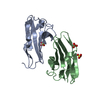 1f5wC 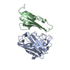 1kacS C: citing same article ( S: Starting model for refinement |
|---|---|
| Similar structure data |


-Links
 PDBj
PDBj


- Assembly
| Deposited unit | 
| ||||||||
|---|---|---|---|---|---|---|---|---|---|
| 1 |
| ||||||||
| Unit cell |
| ||||||||
| Noncrystallographic symmetry (NCS) | NCS oper: (Code: given Matrix: (0.70567, -0.70834, 0.01682), Vector: Details | ANALYTICAL ULTRACENTIFUGATION EXPERIMENTS PROVETHE EXISTENCE OF A DIMER ALSO IN SOLUTION, WITH ADISSOCIATION CONSTANT IN THE MICROMOLAR RANGE.SEE JRNL REFERENCE | |
-Components
| #1: Protein | Mass: 14048.972 Da / Num. of mol.: 2 / Fragment: D1 DOMAIN RESIDUES 15-140 Source method: isolated from a genetically manipulated source Details: DISULFIDE BOND BETWEEN A41 AND A120 AND BETWEEN B41 AND B120 Source: (gene. exp.) HOMO SAPIENS (human) / Description: HELA CELL CDNA LIBRARY / Plasmid: PAB3 / Production host:  #2: Chemical |   Mass: 96.063 Da / Num. of mol.: 2 / Source method: obtained synthetically / Formula: SO4 Mass: 96.063 Da / Num. of mol.: 2 / Source method: obtained synthetically / Formula: SO4#3: Water | ChemComp-HOH / |  Mass: 18.015 Da / Num. of mol.: 332 / Source method: isolated from a natural source / Formula: H2O Mass: 18.015 Da / Num. of mol.: 332 / Source method: isolated from a natural source / Formula: H2OCompound details | RECEPTOR FOR GROUP B COXSACKIEV | Has protein modification | Y | |
|---|
-Experimental details
-Experiment
| Experiment | Method: X-RAY DIFFRACTION / Number of used crystals: 1 |
|---|
- Sample preparation
| Crystal | Density Matthews: 3.1 Å3/Da / Density % sol: 59.5 % |
|---|---|
| Crystal grow | pH: 5.6 Details: AMMONIUM SULPHATE, SODIUM CITRATE, GLYCEROL, pH 5.60 |
| Crystal grow | *PLUS Method: unknown |
-Data collection
| Diffraction | Mean temperature: 100 K |
|---|---|
| Diffraction source | Source: SYNCHROTRON / Site: ESRF  / Beamline: ID14-2 / Wavelength: 0.933 / Beamline: ID14-2 / Wavelength: 0.933 |
| Detector | Type: ADSC 2X2 / Detector: CCD / Date: Jun 1, 2000 |
| Radiation | Protocol: SINGLE WAVELENGTH / Monochromatic (M) / Laue (L): M / Scattering type: x-ray |
| Radiation wavelength | Wavelength: 0.933 Å / Relative weight: 1 |
| Reflection | Resolution: 1.35→20 Å / Num. obs: 73582 / % possible obs: 95.1 % / Redundancy: 4 % / Rmerge(I) obs: 0.063 / Net I/σ(I): 5.9 |
| Reflection shell | Resolution: 1.35→1.42 Å / Redundancy: 1.7 % / Rmerge(I) obs: 0.267 / Mean I/σ(I) obs: 2.1 / % possible all: 68.7 |
- Processing
| Software |
| ||||||||||||||||||||
|---|---|---|---|---|---|---|---|---|---|---|---|---|---|---|---|---|---|---|---|---|---|
| Refinement | Method to determine structure: MOLECULAR REPLACEMENT Starting model: 1KAC, CHAIN B Resolution: 1.35→19 Å / SU B: 1.73 / SU ML: 0.037 / Cross valid method: THROUGHOUT / ESU R Free: 0.036 Details: HYDROGENS HAVE BEEN ADDED IN THE RIDING POSITIONS REFLECTIONS FOR RFREE WERE SELECTED IN THIN SHELLS OF RESOLUTIO, INDEPENDENTLY FROM REFLECTIONS SELECTED FOR RFREE USED IN THE 1.7 ANGSTROM ...Details: HYDROGENS HAVE BEEN ADDED IN THE RIDING POSITIONS REFLECTIONS FOR RFREE WERE SELECTED IN THIN SHELLS OF RESOLUTIO, INDEPENDENTLY FROM REFLECTIONS SELECTED FOR RFREE USED IN THE 1.7 ANGSTROM STRUCTURE. TO ELIMINATE POSSIBLE BIAS FROM THE 1.7 ANGSTROM STRUCTURE, STRUCTURE SOLUTION WAS COMPLETELY REDONE, PLACING TWO COPIES OF CHAIN B FROM PDB-ENTRY 1KAC WITH THE PROGRAM AMORE
| ||||||||||||||||||||
| Refinement step | Cycle: LAST / Resolution: 1.35→19 Å
|
