- PDB-1xl3: Complex structure of Y.pestis virulence Factors YopN and TyeA -
+
Open data
ID or keywords:
Loading...
-
Basic information
Entry
Database: PDB / ID: 1xl3
Title
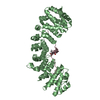
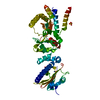
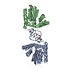
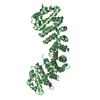
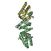
Complex structure of Y.pestis virulence Factors YopN and TyeA
Components
Secretion control protein
protein type A
Keywords
CELL INVASION / yopN / TyeA / Yersinia pestis / type III secretion
Function / homology
Function and homology information
protein secretion by the type III secretion system / negative regulation of protein secretion / cell outer membrane / cell surface Similarity search - Function
Monooxygenase - #80 / Type III secretion system effector delivery regulator TyeA / TyeA / TyeA superfamily / Type III secretion system effector delivery regulator TyeA-related / Type III secretion regulator, YopN/LcrE/InvE/MxiC / Hypersensitivity response secretion-like, HrpJ / HrpJ-like domain / Monooxygenase / Up-down Bundle / Mainly Alpha Similarity search - Domain/homology
#1: Journal: Acta Crystallogr.,Sect.D / Year: 2004 Title: A pivotal role for reductive methylation in the de novo crystallization of a ternary complex composed of Yersinia pestis virulence factors YopN, SycN and YscB Authors: Schubot, F.D. / Waugh, D.S.
In the structure databanks used in Yorodumi, some data are registered as the other names, "COVID-19 virus" and "2019-nCoV". Here are the details of the virus and the list of structure data.
Jan 31, 2019. EMDB accession codes are about to change! (news from PDBe EMDB page)
EMDB accession codes are about to change! (news from PDBe EMDB page)
The allocation of 4 digits for EMDB accession codes will soon come to an end. Whilst these codes will remain in use, new EMDB accession codes will include an additional digit and will expand incrementally as the available range of codes is exhausted. The current 4-digit format prefixed with “EMD-” (i.e. EMD-XXXX) will advance to a 5-digit format (i.e. EMD-XXXXX), and so on. It is currently estimated that the 4-digit codes will be depleted around Spring 2019, at which point the 5-digit format will come into force.
The EM Navigator/Yorodumi systems omit the EMD- prefix.
Related info.:Q: What is EMD? / ID/Accession-code notation in Yorodumi/EM Navigator
Yorodumi is a browser for structure data from EMDB, PDB, SASBDB, etc.
This page is also the successor to EM Navigator detail page, and also detail information page/front-end page for Omokage search.
The word "yorodu" (or yorozu) is an old Japanese word meaning "ten thousand". "mi" (miru) is to see.
Related info.:EMDB / PDB / SASBDB / Comparison of 3 databanks / Yorodumi Search / Aug 31, 2016. New EM Navigator & Yorodumi / Yorodumi Papers / Jmol/JSmol / Function and homology information / Changes in new EM Navigator and Yorodumi
 Movie
Movie Controller


 Open data
 Basic information
 Components
 Keywords
 Function and homology information
 Yersinia pestis (bacteria)
 X-RAY DIFFRACTION /
  Authors
 Citation
 Structure visualization
 Downloads & links
 Other downloads


 PDBj
PDBj Assembly
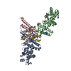

 Mass: 18.015 Da / Num. of mol.: 180 / Source method: isolated from a natural source / Formula: H2O
 Sample preparation
 / Beamline: 22-ID / Wavelength: 1 Å
 Processing