 Movie
Movie Controller

[English] 日本語
 Yorodumi
- PDB-1rj9: Structure of the heterodimer of the conserved GTPase domains of t... -
+ Open data

- Basic information
| Entry | Database: PDB / ID: 1rj9 | ||||||
|---|---|---|---|---|---|---|---|
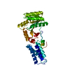
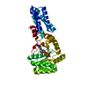
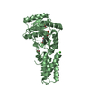
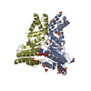
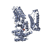
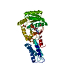
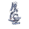
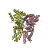
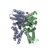
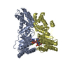
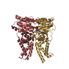
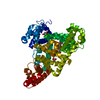
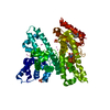
| Title | Structure of the heterodimer of the conserved GTPase domains of the Signal Recognition Particle (Ffh) and Its Receptor (FtsY) | ||||||
 Components Components |
| ||||||
 Keywords Keywords | PROTEIN TRANSPORT / SRP-GTPase DOMAIN / HETERODIMER / NUCLEOTIDE TWINNING / Protein-Protein complex | ||||||
| Function / homology |  Function and homology information Function and homology informationsignal recognition particle / signal recognition particle binding / signal-recognition-particle GTPase / 7S RNA binding / SRP-dependent cotranslational protein targeting to membrane / GTPase activity / GTP binding / plasma membrane / cytoplasm Similarity search - Function | ||||||
| Biological species |   Thermus aquaticus (bacteria) Thermus aquaticus (bacteria) | ||||||
| Method |  X-RAY DIFFRACTION / SYNCHROTRON / MOLECULAR REPLACEMENT / Resolution: 1.9 Å X-RAY DIFFRACTION / SYNCHROTRON / MOLECULAR REPLACEMENT / Resolution: 1.9 Å | ||||||
 Authors Authors | Egea, P.F. / Shan, S.O. / Napetschnig, J. / Savage, D.F. / Walter, P. / Stroud, R.M. | ||||||
 Citation Citation | Journal: Nature / Year: 2004 Title: Substrate twinning activates the signal recognition particle and its receptor Authors: Egea, P.F. / Shan, S.O. / Napetschnig, J. / Savage, D.F. / Walter, P. / Stroud, R.M. | ||||||
| History |
|
- Structure visualization
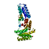
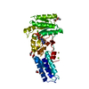
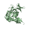
| Structure viewer | Molecule: MolmilJmol/JSmol |
|---|
- Downloads & links
-Download
| PDBx/mmCIF format | 1rj9.cif.gz | 130.6 KB | Display | PDBx/mmCIF format |
|---|---|---|---|---|
| PDB format | pdb1rj9.ent.gz | 99 KB | Display | PDB format |
| PDBx/mmJSON format | 1rj9.json.gz | Tree view | PDBx/mmJSON format | |
| Others |  Other downloads Other downloads |
-Validation report
| Arichive directory | https://data.pdbj.org/pub/pdb/validation_reports/rj/1rj9ftp://data.pdbj.org/pub/pdb/validation_reports/rj/1rj9 | HTTPS FTP |
|---|
-Related structure data
| Related structure data | |
|---|---|
| Similar structure data |


-Links
 PDBj
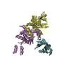
PDBj- Assembly
| Deposited unit | 
| ||||||||
|---|---|---|---|---|---|---|---|---|---|
| 1 |
| ||||||||
| Unit cell |
|
-Components
| #1: Protein | Mass: 33093.387 Da / Num. of mol.: 1 Source method: isolated from a genetically manipulated source Source: (gene. exp.) Thermus aquaticus (bacteria) / Plasmid: pET-28b / Production host: | ||||
|---|---|---|---|---|---|
| #2: Protein | Mass: 32872.977 Da / Num. of mol.: 1 / Fragment: NG domain (Residues 1-300) Source method: isolated from a genetically manipulated source Source: (gene. exp.) Thermus aquaticus (bacteria) / Gene: FFH / Plasmid: pET-28b / Production host: | ||||
| #3: Chemical |   Mass: 24.305 Da / Num. of mol.: 2 / Source method: obtained synthetically / Formula: Mg Mass: 24.305 Da / Num. of mol.: 2 / Source method: obtained synthetically / Formula: Mg#4: Chemical |   Mass: 521.208 Da / Num. of mol.: 2 / Source method: obtained synthetically / Formula: C11H18N5O13P3 / Comment: GMP-PCP, energy-carrying molecule analogue*YM Mass: 521.208 Da / Num. of mol.: 2 / Source method: obtained synthetically / Formula: C11H18N5O13P3 / Comment: GMP-PCP, energy-carrying molecule analogue*YM#5: Water | ChemComp-HOH / |  Mass: 18.015 Da / Num. of mol.: 387 / Source method: isolated from a natural source / Formula: H2O Mass: 18.015 Da / Num. of mol.: 387 / Source method: isolated from a natural source / Formula: H2O |
-Experimental details
-Experiment
| Experiment | Method: X-RAY DIFFRACTION / Number of used crystals: 1 |
|---|
- Sample preparation
| Crystal | Density Matthews: 2.24 Å3/Da / Density % sol: 45 % | ||||||||||||||||||
|---|---|---|---|---|---|---|---|---|---|---|---|---|---|---|---|---|---|---|---|
| Crystal grow | Temperature: 300 K / Method: vapor diffusion, hanging drop / pH: 7 Details: PEG 8000, Hepes, NaCl, pH 7.0, VAPOR DIFFUSION, HANGING DROP, temperature 300K | ||||||||||||||||||
| Crystal grow | *PLUS Temperature: 4 ℃ / pH: 7.5 / Method: vapor diffusion | ||||||||||||||||||
| Components of the solutions | *PLUS
|
-Data collection
| Diffraction | Mean temperature: 100 K |
|---|---|
| Diffraction source | Source: SYNCHROTRON / Site: ALS  / Beamline: 8.3.1 / Wavelength: 1 Å / Beamline: 8.3.1 / Wavelength: 1 Å |
| Radiation | Protocol: SINGLE WAVELENGTH / Monochromatic (M) / Laue (L): M / Scattering type: x-ray |
| Radiation wavelength | Wavelength: 1 Å / Relative weight: 1 |
| Reflection | Resolution: 1.9→50 Å / Num. all: 47344 / Num. obs: 45242 / % possible obs: 95.5 % / Observed criterion σ(F): 0 / Observed criterion σ(I): 0 / Redundancy: 6.3 % / Biso Wilson estimate: 13 Å2 / Rmerge(I) obs: 0.077 / Rsym value: 0.077 / Net I/σ(I): 16.9 |
| Reflection shell | Resolution: 1.9→1.97 Å / Redundancy: 6.3 % / Rmerge(I) obs: 0.77 / Mean I/σ(I) obs: 3.1 / Num. unique all: 4667 / Rsym value: 0.77 / % possible all: 98 |
| Reflection | *PLUS Num. obs: 47344 / % possible obs: 99.3 % / Num. measured all: 305159 |
| Reflection shell | *PLUS % possible obs: 97.9 % / Rmerge(I) obs: 0.767 |
- Processing
| Software |
| |||||||||||||||||||||||||
|---|---|---|---|---|---|---|---|---|---|---|---|---|---|---|---|---|---|---|---|---|---|---|---|---|---|---|
| Refinement | Method to determine structure: MOLECULAR REPLACEMENT Starting model: the structures of isolated FtsY and Ffh NG domain Resolution: 1.9→50 Å / Cross valid method: THROUGHOUT / σ(F): 0 / σ(I): 2 / Stereochemistry target values: Engh & Huber
| |||||||||||||||||||||||||
| Displacement parameters | Biso mean: 39 Å2
| |||||||||||||||||||||||||
| Refinement step | Cycle: LAST / Resolution: 1.9→50 Å
| |||||||||||||||||||||||||
| Refine LS restraints |
| |||||||||||||||||||||||||
| Refinement | *PLUS Num. reflection Rfree: 3436 / % reflection Rfree: 7.5 % | |||||||||||||||||||||||||
| Solvent computation | *PLUS | |||||||||||||||||||||||||
| Displacement parameters | *PLUS | |||||||||||||||||||||||||
| Refine LS restraints | *PLUS
|
