 Movie
Movie Controller

[English] 日本語
 Yorodumi
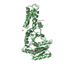
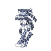
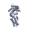
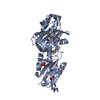
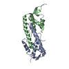
- PDB-2ng1: N AND GTPASE DOMAINS OF THE SIGNAL SEQUENCE RECOGNITION PROTEIN F... -
+ Open data

- Basic information
| Entry | Database: PDB / ID: 2ng1 | ||||||
|---|---|---|---|---|---|---|---|
| Title | N AND GTPASE DOMAINS OF THE SIGNAL SEQUENCE RECOGNITION PROTEIN FFH FROM THERMUS AQUATICUS | ||||||
 Components Components | SIGNAL SEQUENCE RECOGNITION PROTEIN FFH | ||||||
 Keywords Keywords | SIGNAL RECOGNITION / FFH / SRP / GTPASE / SIGNAL RECOGNITION PARTICLE / GDP | ||||||
| Function / homology |  Function and homology information Function and homology informationsignal recognition particle / signal-recognition-particle GTPase / 7S RNA binding / SRP-dependent cotranslational protein targeting to membrane / GTPase activity / GTP binding Similarity search - Function | ||||||
| Biological species |   Thermus aquaticus (bacteria) Thermus aquaticus (bacteria) | ||||||
| Method |  X-RAY DIFFRACTION / MOLECULAR REPLACEMENT / Resolution: 2.02 Å X-RAY DIFFRACTION / MOLECULAR REPLACEMENT / Resolution: 2.02 Å | ||||||
 Authors Authors | Freymann, D.M. / Stroud, R.M. / Walter, P. | ||||||
 Citation Citation | Journal: Nat.Struct.Biol. / Year: 1999 Title: Functional changes in the structure of the SRP GTPase on binding GDP and Mg2+GDP. Authors: Freymann, D.M. / Keenan, R.J. / Stroud, R.M. / Walter, P. #1: Journal: Cell(Cambridge,Mass.) / Year: 1998Title: Crystal Structure of the Signal Sequence Binding Subunit of the Signal Recognition Particle Authors: Keenan, R.J. / Freymann, D.M. / Walter, P. / Stroud, R.M. #2: Journal: Nature / Year: 1997Title: Structure of the Conserved Gtpase Domain of the Signal Recognition Particle Authors: Freymann, D.M. / Keenan, R.J. / Stroud, R.M. / Walter, P. | ||||||
| History |
|
- Structure visualization
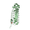
| Structure viewer | Molecule: MolmilJmol/JSmol |
|---|
- Downloads & links
-Download
| PDBx/mmCIF format | 2ng1.cif.gz | 72.2 KB | Display | PDBx/mmCIF format |
|---|---|---|---|---|
| PDB format | pdb2ng1.ent.gz | 52.3 KB | Display | PDB format |
| PDBx/mmJSON format | 2ng1.json.gz | Tree view | PDBx/mmJSON format | |
| Others |  Other downloads Other downloads |
-Validation report
| Arichive directory | https://data.pdbj.org/pub/pdb/validation_reports/ng/2ng1ftp://data.pdbj.org/pub/pdb/validation_reports/ng/2ng1 | HTTPS FTP |
|---|
-Related structure data
| Related structure data | 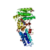 1ng1C 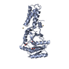 3ng1C 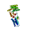 1ffhS S: Starting model for refinement C: citing same article ( |
|---|---|
| Similar structure data |


-Links
 PDBj
PDBj- Assembly
| Deposited unit | 
| ||||||||
|---|---|---|---|---|---|---|---|---|---|
| 1 |
| ||||||||
| Unit cell |
|
-Components
| #1: Protein | Mass: 32199.199 Da / Num. of mol.: 1 / Fragment: NG GTPASE FRAGMENT / Mutation: YES Source method: isolated from a genetically manipulated source Source: (gene. exp.) Thermus aquaticus (bacteria) / Gene: FFH / Plasmid: PET3C / Production host: | ||
|---|---|---|---|
| #2: Chemical | ChemComp-GDP /   Type: RNA linking / Mass: 443.201 Da / Num. of mol.: 1 / Source method: obtained synthetically / Formula: C10H15N5O11P2 / Comment: GDP, energy-carrying molecule*YM Type: RNA linking / Mass: 443.201 Da / Num. of mol.: 1 / Source method: obtained synthetically / Formula: C10H15N5O11P2 / Comment: GDP, energy-carrying molecule*YM | ||
| #3: Chemical | ChemComp-DIO /   Mass: 88.105 Da / Num. of mol.: 1 / Source method: obtained synthetically / Formula: C4H8O2 Mass: 88.105 Da / Num. of mol.: 1 / Source method: obtained synthetically / Formula: C4H8O2 | ||
| #4: Chemical |   Mass: 62.068 Da / Num. of mol.: 2 / Source method: obtained synthetically / Formula: C2H6O2 Mass: 62.068 Da / Num. of mol.: 2 / Source method: obtained synthetically / Formula: C2H6O2#5: Water | ChemComp-HOH / |  Mass: 18.015 Da / Num. of mol.: 72 / Source method: isolated from a natural source / Formula: H2O Mass: 18.015 Da / Num. of mol.: 72 / Source method: isolated from a natural source / Formula: H2O |
-Experimental details
-Experiment
| Experiment | Method: X-RAY DIFFRACTION / Number of used crystals: 1 |
|---|
- Sample preparation
| Crystal | Density Matthews: 2.36 Å3/Da / Density % sol: 47.91 % | ||||||||||||||||||||
|---|---|---|---|---|---|---|---|---|---|---|---|---|---|---|---|---|---|---|---|---|---|
| Crystal grow | *PLUS Method: vapor diffusion, sitting drop | ||||||||||||||||||||
| Components of the solutions | *PLUS
|
-Data collection
| Diffraction | Mean temperature: 100 K |
|---|---|
| Diffraction source | Source: ROTATING ANODE / Type: RIGAKU / Wavelength: 1.5418 |
| Detector | Type: RIGAKU RAXIS / Detector: IMAGE PLATE / Date: Dec 12, 1996 / Details: YALE MIRRORS |
| Radiation | Protocol: SINGLE WAVELENGTH / Monochromatic (M) / Laue (L): M / Scattering type: x-ray |
| Radiation wavelength | Wavelength: 1.5418 Å / Relative weight: 1 |
| Reflection | Resolution: 2.02→20 Å / Num. obs: 18481 / % possible obs: 93 % / Observed criterion σ(I): -3 / Redundancy: 3.2 % / Rmerge(I) obs: 0.072 / Rsym value: 0.072 / Net I/σ(I): 12.9 |
| Reflection shell | Resolution: 2.02→2.09 Å / Redundancy: 2.9 % / Rmerge(I) obs: 0.284 / Mean I/σ(I) obs: 3.6 / Rsym value: 0.284 / % possible all: 93 |
| Reflection shell | *PLUS % possible obs: 92.8 % |
- Processing
| Software |
| ||||||||||||||||||||||||||||||||||||||||||||||||||||||||||||
|---|---|---|---|---|---|---|---|---|---|---|---|---|---|---|---|---|---|---|---|---|---|---|---|---|---|---|---|---|---|---|---|---|---|---|---|---|---|---|---|---|---|---|---|---|---|---|---|---|---|---|---|---|---|---|---|---|---|---|---|---|---|
| Refinement | Method to determine structure: MOLECULAR REPLACEMENT Starting model: PDB ENTRY 1FFH Resolution: 2.02→20 Å / Data cutoff high absF: 10000000 / Data cutoff low absF: 0.1 / Isotropic thermal model: RESTRAINED / Cross valid method: THROUGHOUT / σ(F): 1 Details: A BULK SOLVENT CORRECTION WAS APPLIED. THE DATA QUALITY AND COMPLETENESS BEYOND 2.3 A WERE AFFECTED BY A STRONG ICE RING. THE ELECTRON DENSITY MAPS WERE SOMEWHAT NOISIER THAN WOULD BE ...Details: A BULK SOLVENT CORRECTION WAS APPLIED. THE DATA QUALITY AND COMPLETENESS BEYOND 2.3 A WERE AFFECTED BY A STRONG ICE RING. THE ELECTRON DENSITY MAPS WERE SOMEWHAT NOISIER THAN WOULD BE EXPECTED FOR A STRUCTURE AT THIS RESOLUTION. SEVERAL MAIN CHAIN ATOMS OF THE FOLLOWING SURFACE EXPOSED STRETCHES ARE POORLY DEFINED IN THE ELECTRON DENSITY MAP: 1) PHE 2 - GLN 3, 2) GLY 18 - GLU 25, 3) GLN 63 - GLU 66, AND 4) ALA 251 - ARG 252. THEY ARE CHARACTERIZED BY HIGH TEMPERATURE FACTORS AND ARE PROBABLY MOBILE.
| ||||||||||||||||||||||||||||||||||||||||||||||||||||||||||||
| Displacement parameters | Biso mean: 41 Å2 | ||||||||||||||||||||||||||||||||||||||||||||||||||||||||||||
| Refinement step | Cycle: LAST / Resolution: 2.02→20 Å
| ||||||||||||||||||||||||||||||||||||||||||||||||||||||||||||
| Refine LS restraints |
| ||||||||||||||||||||||||||||||||||||||||||||||||||||||||||||
| LS refinement shell | Resolution: 2.02→2.08 Å / Total num. of bins used: 12
| ||||||||||||||||||||||||||||||||||||||||||||||||||||||||||||
| Xplor file | Serial no: 1 / Param file: PROTEIN_REP.PARAM / Topol file: TOPHCSDX.PRO | ||||||||||||||||||||||||||||||||||||||||||||||||||||||||||||
| Software | *PLUS Name: X-PLOR / Version: 3.851 / Classification: refinement | ||||||||||||||||||||||||||||||||||||||||||||||||||||||||||||
| Refinement | *PLUS Num. reflection obs: 16898 / Num. reflection Rfree: 1320 / Rfactor Rfree: 0.291 | ||||||||||||||||||||||||||||||||||||||||||||||||||||||||||||
| Solvent computation | *PLUS | ||||||||||||||||||||||||||||||||||||||||||||||||||||||||||||
| Displacement parameters | *PLUS | ||||||||||||||||||||||||||||||||||||||||||||||||||||||||||||
| Refine LS restraints | *PLUS
|
