 Movie
Movie Controller

[English] 日本語
 Yorodumi
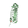
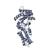
- PDB-3ng1: N AND GTPASE DOMAINS OF THE SIGNAL SEQUENCE RECOGNITION PROTEIN F... -
+ Open data

- Basic information
| Entry | Database: PDB / ID: 3ng1 | ||||||
|---|---|---|---|---|---|---|---|
| Title | N AND GTPASE DOMAINS OF THE SIGNAL SEQUENCE RECOGNITION PROTEIN FFH FROM THERMUS AQUATICUS | ||||||
 Components Components | SIGNAL SEQUENCE RECOGNITION PROTEIN FFH | ||||||
 Keywords Keywords | SIGNAL RECOGNITION / FFH / SRP / GTPASE / SIGNAL RECOGNITION PARTICLE | ||||||
| Function / homology |  Function and homology information Function and homology informationsignal recognition particle / signal-recognition-particle GTPase / 7S RNA binding / SRP-dependent cotranslational protein targeting to membrane / GTPase activity / GTP binding Similarity search - Function | ||||||
| Biological species |   Thermus aquaticus (bacteria) Thermus aquaticus (bacteria) | ||||||
| Method |  X-RAY DIFFRACTION / SYNCHROTRON / MOLECULAR REPLACEMENT / Resolution: 2.3 Å X-RAY DIFFRACTION / SYNCHROTRON / MOLECULAR REPLACEMENT / Resolution: 2.3 Å | ||||||
 Authors Authors | Freymann, D.M. / Stroud, R.M. / Walter, P. | ||||||
 Citation Citation | Journal: Nat.Struct.Biol. / Year: 1999 Title: Functional changes in the structure of the SRP GTPase on binding GDP and Mg2+GDP. Authors: Freymann, D.M. / Keenan, R.J. / Stroud, R.M. / Walter, P. #1: Journal: Cell(Cambridge,Mass.) / Year: 1998Title: Crystal Structure of the Signal Sequence Binding Subunit of the Signal Recognition Particle Authors: Keenan, R.J. / Freymann, D.M. / Walter, P. / Stroud, R.M. #2: Journal: Nature / Year: 1997Title: Structure of the Conserved Gtpase Domain of the Signal Recognition Particle Authors: Freymann, D.M. / Keenan, R.J. / Stroud, R.M. / Walter, P. | ||||||
| History |
|
- Structure visualization
| Structure viewer | Molecule: MolmilJmol/JSmol |
|---|
- Downloads & links
-Download
| PDBx/mmCIF format | 3ng1.cif.gz | 127.6 KB | Display | PDBx/mmCIF format |
|---|---|---|---|---|
| PDB format | pdb3ng1.ent.gz | 100 KB | Display | PDB format |
| PDBx/mmJSON format | 3ng1.json.gz | Tree view | PDBx/mmJSON format | |
| Others |  Other downloads Other downloads |
-Validation report
| Arichive directory | https://data.pdbj.org/pub/pdb/validation_reports/ng/3ng1ftp://data.pdbj.org/pub/pdb/validation_reports/ng/3ng1 | HTTPS FTP |
|---|
-Related structure data
| Related structure data | 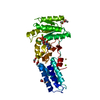 1ng1C 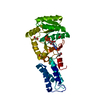 2ng1C 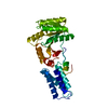 1ffhS S: Starting model for refinement C: citing same article ( |
|---|---|
| Similar structure data |


-Links
 PDBj
PDBj- Assembly
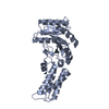
| Deposited unit | 
| ||||||||
|---|---|---|---|---|---|---|---|---|---|
| 1 | 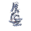
| ||||||||
| 2 | 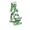
| ||||||||
| Unit cell |
| ||||||||
| Noncrystallographic symmetry (NCS) | NCS oper: (Code: given Matrix: (1, 0.0017, -0.0006), Vector: |
-Components
| #1: Protein | Mass: 32300.371 Da / Num. of mol.: 2 / Fragment: NG GTPASE FRAGMENT Source method: isolated from a genetically manipulated source Source: (gene. exp.) Thermus aquaticus (bacteria) / Gene: FFH / Plasmid: PET3C / Production host: #2: Chemical | ChemComp-CD /   Mass: 112.411 Da / Num. of mol.: 6 / Source method: obtained synthetically / Formula: Cd Mass: 112.411 Da / Num. of mol.: 6 / Source method: obtained synthetically / Formula: Cd#3: Chemical |   Mass: 96.063 Da / Num. of mol.: 2 / Source method: obtained synthetically / Formula: SO4 Mass: 96.063 Da / Num. of mol.: 2 / Source method: obtained synthetically / Formula: SO4#4: Chemical | ChemComp-EDO /   Mass: 62.068 Da / Num. of mol.: 4 / Source method: obtained synthetically / Formula: C2H6O2 Mass: 62.068 Da / Num. of mol.: 4 / Source method: obtained synthetically / Formula: C2H6O2#5: Water | ChemComp-HOH / |  Mass: 18.015 Da / Num. of mol.: 196 / Source method: isolated from a natural source / Formula: H2O Mass: 18.015 Da / Num. of mol.: 196 / Source method: isolated from a natural source / Formula: H2ONonpolymer details | THE CADMIUM ION WHICH MEDIATES THE CRYSTAL CONTACT (CD 701) IS COORDINATED BY GLU 11 AND GLU 33 OF ...THE CADMIUM ION WHICH MEDIATES THE CRYSTAL CONTACT (CD 701) IS COORDINATE | |
|---|
-Experimental details
-Experiment
| Experiment | Method: X-RAY DIFFRACTION / Number of used crystals: 1 |
|---|
- Sample preparation
| Crystal | Density Matthews: 2.8 Å3/Da / Density % sol: 56.12 % | ||||||||||||||||||||||||||||||||||||
|---|---|---|---|---|---|---|---|---|---|---|---|---|---|---|---|---|---|---|---|---|---|---|---|---|---|---|---|---|---|---|---|---|---|---|---|---|---|
| Crystal grow | pH: 6.5 / Details: pH 6.50 | ||||||||||||||||||||||||||||||||||||
| Crystal grow | *PLUS Method: vapor diffusion, sitting drop | ||||||||||||||||||||||||||||||||||||
| Components of the solutions | *PLUS
|
-Data collection
| Diffraction | Mean temperature: 100 K |
|---|---|
| Diffraction source | Source: SYNCHROTRON / Site: SSRL  / Beamline: BL7-1 / Wavelength: 1.08 / Beamline: BL7-1 / Wavelength: 1.08 |
| Detector | Type: MAR scanner 180 mm plate / Detector: IMAGE PLATE / Date: Jul 5, 1997 / Details: MIRROR |
| Radiation | Monochromator: SI(111) / Protocol: SINGLE WAVELENGTH / Monochromatic (M) / Laue (L): M / Scattering type: x-ray |
| Radiation wavelength | Wavelength: 1.08 Å / Relative weight: 1 |
| Reflection | Resolution: 2.3→20 Å / Num. obs: 32541 / % possible obs: 99 % / Observed criterion σ(I): -3 / Redundancy: 4.7 % / Rmerge(I) obs: 0.072 / Rsym value: 0.072 / Net I/σ(I): 14.9 |
| Reflection shell | Resolution: 2.3→2.38 Å / Redundancy: 4.6 % / Rmerge(I) obs: 0.417 / Mean I/σ(I) obs: 3.3 / Rsym value: 0.417 / % possible all: 98 |
| Reflection | *PLUS % possible obs: 98.6 % |
| Reflection shell | *PLUS % possible obs: 98.6 % |
- Processing
| Software |
| ||||||||||||||||||||||||||||||||||||||||||||||||||||||||||||
|---|---|---|---|---|---|---|---|---|---|---|---|---|---|---|---|---|---|---|---|---|---|---|---|---|---|---|---|---|---|---|---|---|---|---|---|---|---|---|---|---|---|---|---|---|---|---|---|---|---|---|---|---|---|---|---|---|---|---|---|---|---|
| Refinement | Method to determine structure: MOLECULAR REPLACEMENT Starting model: PDB ENTRY 1FFH Resolution: 2.3→20 Å / Data cutoff high absF: 10000000 / Data cutoff low absF: 0.1 / Isotropic thermal model: RESTRAINED / Cross valid method: THROUGHOUT / σ(F): 1 / Details: A BULK SOLVENT CORRECTION WAS APPLIED.
| ||||||||||||||||||||||||||||||||||||||||||||||||||||||||||||
| Displacement parameters | Biso mean: 35.4 Å2 | ||||||||||||||||||||||||||||||||||||||||||||||||||||||||||||
| Refinement step | Cycle: LAST / Resolution: 2.3→20 Å
| ||||||||||||||||||||||||||||||||||||||||||||||||||||||||||||
| Refine LS restraints |
| ||||||||||||||||||||||||||||||||||||||||||||||||||||||||||||
| Refine LS restraints NCS | NCS model details: CONSTRAINTS | ||||||||||||||||||||||||||||||||||||||||||||||||||||||||||||
| LS refinement shell | Resolution: 2.3→2.38 Å / Total num. of bins used: 10
| ||||||||||||||||||||||||||||||||||||||||||||||||||||||||||||
| Xplor file | Serial no: 1 / Param file: PROTEIN_REP.PARAM / Topol file: TOPHCSDX.PRO | ||||||||||||||||||||||||||||||||||||||||||||||||||||||||||||
| Software | *PLUS Name: X-PLOR / Version: 3.851 / Classification: refinement | ||||||||||||||||||||||||||||||||||||||||||||||||||||||||||||
| Refine LS restraints | *PLUS
|
