 Movie
Movie Controller


+ Open data

- Basic information
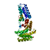
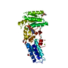
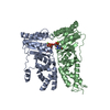
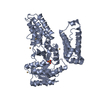
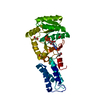
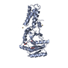
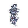
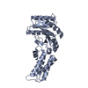
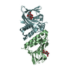
| Entry | Database: PDB / ID: 2c03 | ||||||
|---|---|---|---|---|---|---|---|
| Title | GDP COMPLEX OF SRP GTPASE FFH NG DOMAIN | ||||||
 Components Components | SIGNAL RECOGNITION PARTICLE PROTEIN | ||||||
 Keywords Keywords | SIGNALING PROTEIN / FFH / GMPPNP / GTP-BINDING / NG DOMAIN / RNA-BINDING / SRP54 NUCLEOTIDE-BINDING / SIGNAL PROTEIN | ||||||
| Function / homology |  Function and homology information Function and homology informationsignal recognition particle / signal-recognition-particle GTPase / 7S RNA binding / SRP-dependent cotranslational protein targeting to membrane / GTPase activity / GTP binding Similarity search - Function | ||||||
| Biological species |   THERMUS AQUATICUS (bacteria) THERMUS AQUATICUS (bacteria) | ||||||
| Method |  X-RAY DIFFRACTION / SYNCHROTRON / MOLECULAR REPLACEMENT / Resolution: 1.24 Å X-RAY DIFFRACTION / SYNCHROTRON / MOLECULAR REPLACEMENT / Resolution: 1.24 Å | ||||||
 Authors Authors | Ramirez, U.D. / Preininger, A.M. / Freymann, D.M. | ||||||
 Citation Citation | Journal: Acta Crystallogr.,Sect.D / Year: 2008 Title: Nucleotide-Binding Flexibility in Ultrahigh-Resolution Structures of the Srp Gtpase Ffh Authors: Ramirez, U.D. / Focia, P.J. / Freymann, D.M. | ||||||
| History |
|
- Structure visualization
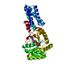
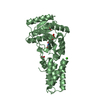
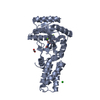
| Structure viewer | Molecule: MolmilJmol/JSmol |
|---|
- Downloads & links
-Download
| PDBx/mmCIF format | 2c03.cif.gz | 606.4 KB | Display | PDBx/mmCIF format |
|---|---|---|---|---|
| PDB format | pdb2c03.ent.gz | 512.4 KB | Display | PDB format |
| PDBx/mmJSON format | 2c03.json.gz | Tree view | PDBx/mmJSON format | |
| Others |  Other downloads Other downloads |
-Validation report
| Arichive directory | https://data.pdbj.org/pub/pdb/validation_reports/c0/2c03ftp://data.pdbj.org/pub/pdb/validation_reports/c0/2c03 | HTTPS FTP |
|---|
-Related structure data
| Related structure data | 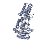 2c04C 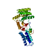 1ffhS  2bqs 2bqt S: Starting model for refinement C: citing same article ( |
|---|---|
| Similar structure data |


-Links
 PDBj
PDBj- Assembly
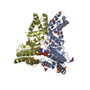
| Deposited unit | 
| ||||||||
|---|---|---|---|---|---|---|---|---|---|
| 1 | 
| ||||||||
| 2 | 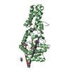
| ||||||||
| Unit cell |
| ||||||||
| Noncrystallographic symmetry (NCS) | NCS oper: (Code: given Matrix: (0.873394, 0.008997, -0.486931), Vector: |
-Components
| #1: Protein | Mass: 32601.660 Da / Num. of mol.: 2 / Fragment: NG, RESIDUES 1-296 Source method: isolated from a genetically manipulated source Source: (gene. exp.) THERMUS AQUATICUS (bacteria) / Production host: #2: Chemical |   Type: RNA linking / Mass: 443.201 Da / Num. of mol.: 2 / Source method: obtained synthetically / Formula: C10H15N5O11P2 / Comment: GDP, energy-carrying molecule*YM Type: RNA linking / Mass: 443.201 Da / Num. of mol.: 2 / Source method: obtained synthetically / Formula: C10H15N5O11P2 / Comment: GDP, energy-carrying molecule*YM#3: Chemical |   Mass: 62.068 Da / Num. of mol.: 3 / Source method: obtained synthetically / Formula: C2H6O2 Mass: 62.068 Da / Num. of mol.: 3 / Source method: obtained synthetically / Formula: C2H6O2#4: Chemical | ChemComp-DIO /   Mass: 88.105 Da / Num. of mol.: 14 / Source method: obtained synthetically / Formula: C4H8O2 Mass: 88.105 Da / Num. of mol.: 14 / Source method: obtained synthetically / Formula: C4H8O2#5: Water | ChemComp-HOH / |  Mass: 18.015 Da / Num. of mol.: 631 / Source method: isolated from a natural source / Formula: H2O Mass: 18.015 Da / Num. of mol.: 631 / Source method: isolated from a natural source / Formula: H2OCompound details | MANDATORY FOR EFFICIENT EXPORT OF EXTRA-CYTOPLASMI | |
|---|
-Experimental details
-Experiment
| Experiment | Method: X-RAY DIFFRACTION / Number of used crystals: 1 |
|---|
- Sample preparation
| Crystal | Density Matthews: 1.5 Å3/Da / Density % sol: 43.9 % Description: DATA WERE COLLECTED IN TWO OVERLAPPING RESOLUTION RANGES |
|---|---|
| Crystal grow | Method: vapor diffusion, hanging drop / Details: 30% DIOXANE, 2MM GDP |
-Data collection
| Diffraction | Mean temperature: 100 K |
|---|---|
| Diffraction source | Source: SYNCHROTRON / Site: APS  / Beamline: 5ID-B / Wavelength: 0.7429 / Beamline: 5ID-B / Wavelength: 0.7429 |
| Detector | Type: MARRESEARCH / Detector: CCD / Date: Sep 14, 1998 |
| Radiation | Protocol: SINGLE WAVELENGTH / Monochromatic (M) / Laue (L): M / Scattering type: x-ray |
| Radiation wavelength | Wavelength: 0.7429 Å / Relative weight: 1 |
| Reflection | Resolution: 1.24→50 Å / Num. obs: 154602 / % possible obs: 94.6 % / Observed criterion σ(I): 3 / Redundancy: 3.6 % / Rmerge(I) obs: 0.063 / Net I/σ(I): 12.5 |
| Reflection shell | Resolution: 1.24→1.27 Å / Redundancy: 3 % / Rmerge(I) obs: 0.47 / Mean I/σ(I) obs: 2 / % possible all: 96.1 |
- Processing
| Software |
| ||||||||||||||||||||||||||||||||||||||||||||||||||||||||||||||||||||||||||||||||||||||||||||||||||||||||||||||||||||||||||||||||||||||||||||||||||||||||||||||||||||||||||||||||||||||
|---|---|---|---|---|---|---|---|---|---|---|---|---|---|---|---|---|---|---|---|---|---|---|---|---|---|---|---|---|---|---|---|---|---|---|---|---|---|---|---|---|---|---|---|---|---|---|---|---|---|---|---|---|---|---|---|---|---|---|---|---|---|---|---|---|---|---|---|---|---|---|---|---|---|---|---|---|---|---|---|---|---|---|---|---|---|---|---|---|---|---|---|---|---|---|---|---|---|---|---|---|---|---|---|---|---|---|---|---|---|---|---|---|---|---|---|---|---|---|---|---|---|---|---|---|---|---|---|---|---|---|---|---|---|---|---|---|---|---|---|---|---|---|---|---|---|---|---|---|---|---|---|---|---|---|---|---|---|---|---|---|---|---|---|---|---|---|---|---|---|---|---|---|---|---|---|---|---|---|---|---|---|---|---|
| Refinement | Method to determine structure: MOLECULAR REPLACEMENT Starting model: PDB ENTRY 1FFH Resolution: 1.24→19.96 Å / Cor.coef. Fo:Fc: 0.979 / Cor.coef. Fo:Fc free: 0.967 / SU B: 1.852 / SU ML: 0.036 / Cross valid method: THROUGHOUT / ESU R: 0.057 / ESU R Free: 0.057 / Stereochemistry target values: MAXIMUM LIKELIHOOD / Details: HYDROGENS HAVE BEEN ADDED IN THE RIDING POSITIONS.
| ||||||||||||||||||||||||||||||||||||||||||||||||||||||||||||||||||||||||||||||||||||||||||||||||||||||||||||||||||||||||||||||||||||||||||||||||||||||||||||||||||||||||||||||||||||||
| Solvent computation | Ion probe radii: 0.8 Å / Shrinkage radii: 0.8 Å / VDW probe radii: 1.2 Å / Solvent model: BABINET MODEL WITH MASK | ||||||||||||||||||||||||||||||||||||||||||||||||||||||||||||||||||||||||||||||||||||||||||||||||||||||||||||||||||||||||||||||||||||||||||||||||||||||||||||||||||||||||||||||||||||||
| Displacement parameters | Biso mean: 21.53 Å2
| ||||||||||||||||||||||||||||||||||||||||||||||||||||||||||||||||||||||||||||||||||||||||||||||||||||||||||||||||||||||||||||||||||||||||||||||||||||||||||||||||||||||||||||||||||||||
| Refinement step | Cycle: LAST / Resolution: 1.24→19.96 Å
| ||||||||||||||||||||||||||||||||||||||||||||||||||||||||||||||||||||||||||||||||||||||||||||||||||||||||||||||||||||||||||||||||||||||||||||||||||||||||||||||||||||||||||||||||||||||
| Refine LS restraints |
|