 Movie
Movie Controller

[English] 日本語
 Yorodumi
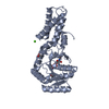
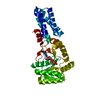
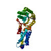
- PDB-1ffh: N AND GTPASE DOMAINS OF THE SIGNAL SEQUENCE RECOGNITION PROTEIN F... -
+ Open data

- Basic information
| Entry | Database: PDB / ID: 1ffh | ||||||
|---|---|---|---|---|---|---|---|
| Title | N AND GTPASE DOMAINS OF THE SIGNAL SEQUENCE RECOGNITION PROTEIN FFH FROM THERMUS AQUATICUS | ||||||
 Components Components | FFH | ||||||
 Keywords Keywords | RIBONUCLEOPROTEIN / FFH / SRP / GTPASE / SIGNAL RECOGNITION PARTICLE | ||||||
| Function / homology |  Function and homology information Function and homology informationsignal recognition particle / signal-recognition-particle GTPase / 7S RNA binding / SRP-dependent cotranslational protein targeting to membrane / GTPase activity / GTP binding Similarity search - Function | ||||||
| Biological species |   Thermus aquaticus (bacteria) Thermus aquaticus (bacteria) | ||||||
| Method |  X-RAY DIFFRACTION / MIR / Resolution: 2.05 Å X-RAY DIFFRACTION / MIR / Resolution: 2.05 Å | ||||||
 Authors Authors | Freymann, D.M. / Keenan, R.J. / Stroud, R.M. / Walter, P. | ||||||
 Citation Citation | Journal: Nature / Year: 1997 Title: Structure of the conserved GTPase domain of the signal recognition particle. Authors: Freymann, D.M. / Keenan, R.J. / Stroud, R.M. / Walter, P. | ||||||
| History |
|
- Structure visualization
| Structure viewer | Molecule: MolmilJmol/JSmol |
|---|
- Downloads & links
-Download
| PDBx/mmCIF format | 1ffh.cif.gz | 69.7 KB | Display | PDBx/mmCIF format |
|---|---|---|---|---|
| PDB format | pdb1ffh.ent.gz | 51.2 KB | Display | PDB format |
| PDBx/mmJSON format | 1ffh.json.gz | Tree view | PDBx/mmJSON format | |
| Others |  Other downloads Other downloads |
-Validation report
| Arichive directory | https://data.pdbj.org/pub/pdb/validation_reports/ff/1ffhftp://data.pdbj.org/pub/pdb/validation_reports/ff/1ffh | HTTPS FTP |
|---|
-Related structure data
| Similar structure data |
|---|


-Links
 PDBj
PDBj- Assembly
| Deposited unit | 
| ||||||||
|---|---|---|---|---|---|---|---|---|---|
| 1 |
| ||||||||
| Unit cell |
|
-Components
| #1: Protein | Mass: 32256.250 Da / Num. of mol.: 1 / Fragment: 'NG' GTPASE FRAGMENT OF FFH Source method: isolated from a genetically manipulated source Details: THE NG FRAGMENT WAS GENERATED BY PROTEOLYSIS / Source: (gene. exp.) Thermus aquaticus (bacteria) / Cell line: BL21 / Gene: FFH / Plasmid: PET3C / Production host: |
|---|---|
| #2: Chemical | ChemComp-MG /   Mass: 24.305 Da / Num. of mol.: 1 / Source method: obtained synthetically / Formula: Mg Mass: 24.305 Da / Num. of mol.: 1 / Source method: obtained synthetically / Formula: Mg |
| #3: Water | ChemComp-HOH /  Mass: 18.015 Da / Num. of mol.: 121 / Source method: isolated from a natural source / Formula: H2O Mass: 18.015 Da / Num. of mol.: 121 / Source method: isolated from a natural source / Formula: H2O |
| Sequence details | FEATURES IN THE FO-FC MAP SUGGEST THAT ALA 48 (BASED ON THE SEQUENCE OF THE T. AQUATICUS FFH GENE) ...FEATURES IN THE FO-FC MAP SUGGEST THAT ALA 48 (BASED ON THE SEQUENCE OF THE T. AQUATICUS FFH GENE) IS THR, SER, OR VAL IN THIS STRUCTURE. THE EXPRESSION |
-Experimental details
-Experiment
| Experiment | Method: X-RAY DIFFRACTION / Number of used crystals: 1 |
|---|
- Sample preparation
| Crystal | Density Matthews: 2.08 Å3/Da / Density % sol: 40.79 % | ||||||||||||||||||||||||||||||
|---|---|---|---|---|---|---|---|---|---|---|---|---|---|---|---|---|---|---|---|---|---|---|---|---|---|---|---|---|---|---|---|
| Crystal grow | Method: vapor diffusion, sitting drop / pH: 9 Details: PROTEIN AT 30 MG/ML IN WATER. CRYSTALLIZED AT RT BY SITTING DROP VAPOR DIFFUSION USING 30% PEG MME 550, 50 MM TAPS PH 9.0, 200 MM MGCL2., vapor diffusion - sitting drop | ||||||||||||||||||||||||||||||
| Crystal grow | *PLUS Method: vapor diffusion, sitting drop | ||||||||||||||||||||||||||||||
| Components of the solutions | *PLUS
|
-Data collection
| Diffraction | Mean temperature: 100 K |
|---|---|
| Diffraction source | Source: ROTATING ANODE / Type: RIGAKU / Wavelength: 1.5418 |
| Detector | Type: MAR scanner 300 mm plate / Detector: IMAGE PLATE / Date: Mar 19, 1996 / Details: SUPPER DOUBLE MIRRORS |
| Radiation | Monochromator: DOUBLE CRYSTAL SI(111) / Monochromatic (M) / Laue (L): M / Scattering type: x-ray |
| Radiation wavelength | Wavelength: 1.5418 Å / Relative weight: 1 |
| Reflection | Highest resolution: 2.05 Å / Num. obs: 16644 / % possible obs: 99.5 % / Observed criterion σ(I): -3 / Redundancy: 4 % / Biso Wilson estimate: 13.2 Å2 / Rsym value: 0.046 |
| Reflection shell | Resolution: 2.05→2.12 Å / Redundancy: 3.9 % / Rsym value: 0.137 / % possible all: 98.4 |
| Reflection | *PLUS Rmerge(I) obs: 0.046 |
| Reflection shell | *PLUS % possible obs: 98.4 % / Rmerge(I) obs: 0.137 |
- Processing
| Software |
| ||||||||||||||||||||||||||||||||||||||||||||||||||||||||||||
|---|---|---|---|---|---|---|---|---|---|---|---|---|---|---|---|---|---|---|---|---|---|---|---|---|---|---|---|---|---|---|---|---|---|---|---|---|---|---|---|---|---|---|---|---|---|---|---|---|---|---|---|---|---|---|---|---|---|---|---|---|---|
| Refinement | Method to determine structure: MIR / Resolution: 2.05→20 Å / Isotropic thermal model: RESTRAINED / Cross valid method: THROUGHOUT / σ(F): 1 Details: THE ELECTRON DENSITY FOR THE TURN COMPRISING THESE RESIDUES IS NOT YET CORRECTLY MODELED: THR 23, GLU 24, GLU 25.
| ||||||||||||||||||||||||||||||||||||||||||||||||||||||||||||
| Displacement parameters | Biso mean: 21.2 Å2 | ||||||||||||||||||||||||||||||||||||||||||||||||||||||||||||
| Refinement step | Cycle: LAST / Resolution: 2.05→20 Å
| ||||||||||||||||||||||||||||||||||||||||||||||||||||||||||||
| Refine LS restraints |
| ||||||||||||||||||||||||||||||||||||||||||||||||||||||||||||
| LS refinement shell | Resolution: 2.05→2.11 Å / Total num. of bins used: 12
| ||||||||||||||||||||||||||||||||||||||||||||||||||||||||||||
| Xplor file |
| ||||||||||||||||||||||||||||||||||||||||||||||||||||||||||||
| Software | *PLUS Name: X-PLOR / Version: 3.843 / Classification: refinement | ||||||||||||||||||||||||||||||||||||||||||||||||||||||||||||
| Refinement | *PLUS | ||||||||||||||||||||||||||||||||||||||||||||||||||||||||||||
| Solvent computation | *PLUS | ||||||||||||||||||||||||||||||||||||||||||||||||||||||||||||
| Displacement parameters | *PLUS | ||||||||||||||||||||||||||||||||||||||||||||||||||||||||||||
| Refine LS restraints | *PLUS
| ||||||||||||||||||||||||||||||||||||||||||||||||||||||||||||
| LS refinement shell | *PLUS Rfactor obs: 0.248 |
