 Movie
Movie Controller


+ Open data

- Basic information
| Entry | Database: PDB / ID: 1ls1 | ||||||
|---|---|---|---|---|---|---|---|
| Title | T. aquaticus Ffh NG Domain at 1.1A Resolution | ||||||
 Components Components | SIGNAL RECOGNITION PARTICLE PROTEIN | ||||||
 Keywords Keywords | PROTEIN TRANSPORT / Ffh / SRP54 / SRP / GTPase / ultrahigh resolution | ||||||
| Function / homology |  Function and homology information Function and homology informationsignal recognition particle / signal-recognition-particle GTPase / 7S RNA binding / SRP-dependent cotranslational protein targeting to membrane / GTPase activity / GTP binding Similarity search - Function | ||||||
| Biological species |   Thermus aquaticus (bacteria) Thermus aquaticus (bacteria) | ||||||
| Method |  X-RAY DIFFRACTION / SYNCHROTRON / MOLECULAR SUBSTITUTION / Resolution: 1.1 Å X-RAY DIFFRACTION / SYNCHROTRON / MOLECULAR SUBSTITUTION / Resolution: 1.1 Å | ||||||
 Authors Authors | Ramirez, U.D. / Minasov, G. / Freymann, D.M. | ||||||
 Citation Citation | Journal: J.Mol.Biol. / Year: 2002 Title: Structural Basis for Mobility in the 1.1 A Crystal Structure of the NG Domain of Thermus aquaticus Ffh Authors: Ramirez, U.D. / Minasov, G. / Focia, P.J. / Stroud, R.M. / Walter, P. / Kuhn, P. / Freymann, D.M. | ||||||
| History |
|
- Structure visualization
| Structure viewer | Molecule: MolmilJmol/JSmol |
|---|
- Downloads & links
-Download
| PDBx/mmCIF format | 1ls1.cif.gz | 202.4 KB | Display | PDBx/mmCIF format |
|---|---|---|---|---|
| PDB format | pdb1ls1.ent.gz | 166.1 KB | Display | PDB format |
| PDBx/mmJSON format | 1ls1.json.gz | Tree view | PDBx/mmJSON format | |
| Others |  Other downloads Other downloads |
-Validation report
| Arichive directory | https://data.pdbj.org/pub/pdb/validation_reports/ls/1ls1ftp://data.pdbj.org/pub/pdb/validation_reports/ls/1ls1 | HTTPS FTP |
|---|
-Related structure data
| Related structure data | 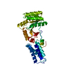 1ffhS S: Starting model for refinement |
|---|---|
| Similar structure data |


-Links
 PDBj
PDBj


- Assembly
| Deposited unit | 
| ||||||||
|---|---|---|---|---|---|---|---|---|---|
| 1 |
| ||||||||
| Unit cell |
| ||||||||
| Components on special symmetry positions |
|
-Components
| #1: Protein | Mass: 32357.422 Da / Num. of mol.: 1 / Fragment: NG domain Source method: isolated from a genetically manipulated source Source: (gene. exp.) Thermus aquaticus (bacteria) / Production host: | ||
|---|---|---|---|
| #2: Chemical | ChemComp-MG /   Mass: 24.305 Da / Num. of mol.: 1 / Source method: obtained synthetically / Formula: Mg Mass: 24.305 Da / Num. of mol.: 1 / Source method: obtained synthetically / Formula: Mg | ||
| #3: Chemical |   Mass: 31.999 Da / Num. of mol.: 3 / Source method: obtained synthetically / Formula: O2 Mass: 31.999 Da / Num. of mol.: 3 / Source method: obtained synthetically / Formula: O2#4: Water | ChemComp-HOH / |  Mass: 18.015 Da / Num. of mol.: 284 / Source method: isolated from a natural source / Formula: H2O Mass: 18.015 Da / Num. of mol.: 284 / Source method: isolated from a natural source / Formula: H2O |
-Experimental details
-Experiment
| Experiment | Method: X-RAY DIFFRACTION / Number of used crystals: 1 |
|---|
- Sample preparation
| Crystal | Density Matthews: 2.07 Å3/Da / Density % sol: 40.65 % | |||||||||||||||||||||||||||||||||||
|---|---|---|---|---|---|---|---|---|---|---|---|---|---|---|---|---|---|---|---|---|---|---|---|---|---|---|---|---|---|---|---|---|---|---|---|---|
| Crystal grow | Temperature: 295 K / Method: vapor diffusion, sitting drop / pH: 9 Details: PEGMME550, MGCl2, TAPS, pH 9.0, VAPOR DIFFUSION, SITTING DROP, temperature 295K | |||||||||||||||||||||||||||||||||||
| Crystal grow | *PLUS | |||||||||||||||||||||||||||||||||||
| Components of the solutions | *PLUS
|
-Data collection
| Diffraction | Mean temperature: 100 K |
|---|---|
| Diffraction source | Source: SYNCHROTRON / Site: SSRL  / Beamline: BL9-1 / Wavelength: 0.98 Å / Beamline: BL9-1 / Wavelength: 0.98 Å |
| Detector | Type: MARRESEARCH / Detector: IMAGE PLATE / Date: Apr 24, 1997 |
| Radiation | Monochromator: SSRL BL 9-1 / Protocol: SINGLE WAVELENGTH / Monochromatic (M) / Laue (L): M / Scattering type: x-ray |
| Radiation wavelength | Wavelength: 0.98 Å / Relative weight: 1 |
| Reflection | Resolution: 1.1→50 Å / Num. obs: 102368 / % possible obs: 95.4 % / Observed criterion σ(F): -3 / Observed criterion σ(I): -3 / Redundancy: 4.27 % / Rmerge(I) obs: 0.037 / Rsym value: 0.037 |
| Reflection shell | Resolution: 1.1→1.12 Å / Redundancy: 2.96 % / Rmerge(I) obs: 0.324 / Mean I/σ(I) obs: 3.2 / Rsym value: 0.324 / % possible all: 90 |
| Reflection | *PLUS Num. measured all: 694235 |
| Reflection shell | *PLUS % possible obs: 89.9 % |
- Processing
| Software |
| ||||||||||||||||||||||||||||||||||||||||||||||||||||||||||||||||||||||||||||||||||||||||||||||||||||||||||||||||||||||||
|---|---|---|---|---|---|---|---|---|---|---|---|---|---|---|---|---|---|---|---|---|---|---|---|---|---|---|---|---|---|---|---|---|---|---|---|---|---|---|---|---|---|---|---|---|---|---|---|---|---|---|---|---|---|---|---|---|---|---|---|---|---|---|---|---|---|---|---|---|---|---|---|---|---|---|---|---|---|---|---|---|---|---|---|---|---|---|---|---|---|---|---|---|---|---|---|---|---|---|---|---|---|---|---|---|---|---|---|---|---|---|---|---|---|---|---|---|---|---|---|---|---|
| Refinement | Method to determine structure: MOLECULAR SUBSTITUTION Starting model: PDB ENTRY 1ffh Resolution: 1.1→50 Å / Cor.coef. Fo:Fc: 0.98 / Cor.coef. Fo:Fc free: 0.972 / SU B: 0.45 / SU ML: 0.021 / Cross valid method: THROUGHOUT / σ(F): 0 / ESU R: 0.032 / ESU R Free: 0.034 / Stereochemistry target values: MAXIMUM LIKELIHOOD / Details: HYDROGENS HAVE BEEN ADDED IN THE RIDING POSITIONS
| ||||||||||||||||||||||||||||||||||||||||||||||||||||||||||||||||||||||||||||||||||||||||||||||||||||||||||||||||||||||||
| Solvent computation | Ion probe radii: 0.8 Å / Shrinkage radii: 0.8 Å / VDW probe radii: 1.4 Å / Solvent model: BABINET MODEL WITH MASK | ||||||||||||||||||||||||||||||||||||||||||||||||||||||||||||||||||||||||||||||||||||||||||||||||||||||||||||||||||||||||
| Displacement parameters | Biso mean: 20.404 Å2 | ||||||||||||||||||||||||||||||||||||||||||||||||||||||||||||||||||||||||||||||||||||||||||||||||||||||||||||||||||||||||
| Refinement step | Cycle: LAST / Resolution: 1.1→50 Å
| ||||||||||||||||||||||||||||||||||||||||||||||||||||||||||||||||||||||||||||||||||||||||||||||||||||||||||||||||||||||||
| Refine LS restraints |
| ||||||||||||||||||||||||||||||||||||||||||||||||||||||||||||||||||||||||||||||||||||||||||||||||||||||||||||||||||||||||
| LS refinement shell | Resolution: 1.1→1.128 Å / Total num. of bins used: 20 /
| ||||||||||||||||||||||||||||||||||||||||||||||||||||||||||||||||||||||||||||||||||||||||||||||||||||||||||||||||||||||||
| Refinement | *PLUS Lowest resolution: 50 Å / % reflection Rfree: 8 % / Rfactor Rfree: 0.169 / Rfactor Rwork: 0.135 | ||||||||||||||||||||||||||||||||||||||||||||||||||||||||||||||||||||||||||||||||||||||||||||||||||||||||||||||||||||||||
| Solvent computation | *PLUS | ||||||||||||||||||||||||||||||||||||||||||||||||||||||||||||||||||||||||||||||||||||||||||||||||||||||||||||||||||||||||
| Displacement parameters | *PLUS | ||||||||||||||||||||||||||||||||||||||||||||||||||||||||||||||||||||||||||||||||||||||||||||||||||||||||||||||||||||||||
| Refine LS restraints | *PLUS
|
