 Movie
Movie Controller


+ Open data

- Basic information
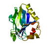
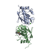
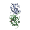
| Entry | Database: PDB / ID: 1cyw | ||||||
|---|---|---|---|---|---|---|---|
| Title | QUINOL OXIDASE (PERIPLASMIC FRAGMENT OF SUBUNIT II) (CYOA) | ||||||
 Components Components | CYOA | ||||||
 Keywords Keywords | ELECTRON TRANSPORT | ||||||
| Function / homology |  Function and homology information Function and homology informationoxidoreduction-driven active transmembrane transporter activity / cytochrome o ubiquinol oxidase complex / cytochrome bo3 ubiquinol oxidase activity / aerobic electron transport chain / respiratory chain complex / oxidoreductase activity, acting on diphenols and related substances as donors, oxygen as acceptor / cytochrome-c oxidase activity / proton transmembrane transporter activity / electron transport coupled proton transport / ATP synthesis coupled electron transport ...oxidoreduction-driven active transmembrane transporter activity / cytochrome o ubiquinol oxidase complex / cytochrome bo3 ubiquinol oxidase activity / aerobic electron transport chain / respiratory chain complex / oxidoreductase activity, acting on diphenols and related substances as donors, oxygen as acceptor / cytochrome-c oxidase activity / proton transmembrane transporter activity / electron transport coupled proton transport / ATP synthesis coupled electron transport / aerobic respiration / electron transfer activity / copper ion binding / plasma membrane Similarity search - Function | ||||||
| Biological species |  | ||||||
| Method |  X-RAY DIFFRACTION / Resolution: 2.5 Å X-RAY DIFFRACTION / Resolution: 2.5 Å | ||||||
 Authors Authors | Wilmanns, M. / Lappalainen, P. / Kelly, M. / Sauer-Eriksson, E. / Saraste, M. | ||||||
 Citation Citation | Journal: Proc.Natl.Acad.Sci.USA / Year: 1995 Title: Crystal structure of the membrane-exposed domain from a respiratory quinol oxidase complex with an engineered dinuclear copper center. Authors: Wilmanns, M. / Lappalainen, P. / Kelly, M. / Sauer-Eriksson, E. / Saraste, M. #1: Journal: J.Mol.Biol. / Year: 1993Title: Crystallization and Preliminary X-Ray Analysis of the Periplasmic Fragment of Cyoa-A Subunit of the Escherichia Coli Cytochrome O Complex Authors: Van Der Oost, J. / Musacchio, A. / Pauptit, R.A. / Ceska, T.A. / Wierenga, R.K. / Saraste, M. #2: Journal: Embo J. / Year: 1992Title: Restoration of a Lost Metal-Binding Site: Construction of Two Different Copper Sites Into a Subunit of the E. Coli Cytochrome O Quinol Oxidase Complex Authors: Van Der Oost, J. / Lappalainen, P. / Musacchio, A. / Warne, A. / Lemieux, L. / Rumbley, J. / Gennis, R.B. / Aasa, R. / Pascher, T. / Malmstrom, B.M. / Saraste, M. | ||||||
| History |
|
- Structure visualization
| Structure viewer | Molecule: MolmilJmol/JSmol |
|---|
- Downloads & links
-Download
| PDBx/mmCIF format | 1cyw.cif.gz | 44.6 KB | Display | PDBx/mmCIF format |
|---|---|---|---|---|
| PDB format | pdb1cyw.ent.gz | 31 KB | Display | PDB format |
| PDBx/mmJSON format | 1cyw.json.gz | Tree view | PDBx/mmJSON format | |
| Others |  Other downloads Other downloads |
-Validation report
| Arichive directory | https://data.pdbj.org/pub/pdb/validation_reports/cy/1cywftp://data.pdbj.org/pub/pdb/validation_reports/cy/1cyw | HTTPS FTP |
|---|
-Related structure data


-Links
 PDBj
PDBj


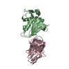
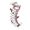
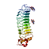
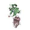
- Assembly
| Deposited unit | 
| ||||||||
|---|---|---|---|---|---|---|---|---|---|
| 1 | 
| ||||||||
| 2 | 
| ||||||||
| Unit cell |
|
-Components
| #1: Protein | Mass: 22703.582 Da / Num. of mol.: 1 / Fragment: PERIPLASMIC FRAGMENT (RESIDUES 111 - 315) Source method: isolated from a genetically manipulated source Source: (gene. exp.) References: UniProt: P0ABJ1, Oxidoreductases; Acting on diphenols and related substances as donors; With oxygen as acceptor |
|---|---|
| #2: Water | ChemComp-HOH /  Mass: 18.015 Da / Num. of mol.: 46 / Source method: isolated from a natural source / Formula: H2O Mass: 18.015 Da / Num. of mol.: 46 / Source method: isolated from a natural source / Formula: H2O |
-Experimental details
-Experiment
| Experiment | Method: X-RAY DIFFRACTION |
|---|
- Sample preparation
| Crystal | Density Matthews: 2.09 Å3/Da / Density % sol: 41.03 % Description: Authors state that several data sets were collected from 1992 to 1994 |
|---|
-Data collection
| Diffraction source | Wavelength: 1.5418 |
|---|---|
| Detector | Type: ENRAF-NONIUS FAST / Detector: DIFFRACTOMETER / Date: 1994 |
| Radiation | Protocol: SINGLE WAVELENGTH / Monochromatic (M) / Laue (L): M / Scattering type: x-ray |
| Radiation wavelength | Wavelength: 1.5418 Å / Relative weight: 1 |
| Reflection | Resolution: 2.5→1000 Å / Num. obs: 5943 / % possible obs: 86.3 % / Observed criterion σ(I): 0 |
| Reflection | *PLUS Num. measured all: 12996 / Rmerge(I) obs: 0.07 |
- Processing
| Software |
| ||||||||||||||||||||||||||||||||||||||||||||||||||||||||||||
|---|---|---|---|---|---|---|---|---|---|---|---|---|---|---|---|---|---|---|---|---|---|---|---|---|---|---|---|---|---|---|---|---|---|---|---|---|---|---|---|---|---|---|---|---|---|---|---|---|---|---|---|---|---|---|---|---|---|---|---|---|---|
| Refinement | Resolution: 2.5→6 Å / σ(F): 1
| ||||||||||||||||||||||||||||||||||||||||||||||||||||||||||||
| Displacement parameters | Biso mean: 32.7 Å2 | ||||||||||||||||||||||||||||||||||||||||||||||||||||||||||||
| Refine analyze | Luzzati coordinate error obs: 0.3 Å | ||||||||||||||||||||||||||||||||||||||||||||||||||||||||||||
| Refinement step | Cycle: LAST / Resolution: 2.5→6 Å
| ||||||||||||||||||||||||||||||||||||||||||||||||||||||||||||
| Refine LS restraints |
| ||||||||||||||||||||||||||||||||||||||||||||||||||||||||||||
| Software | *PLUS Name: X-PLOR / Classification: refinement | ||||||||||||||||||||||||||||||||||||||||||||||||||||||||||||
| Refinement | *PLUS | ||||||||||||||||||||||||||||||||||||||||||||||||||||||||||||
| Solvent computation | *PLUS | ||||||||||||||||||||||||||||||||||||||||||||||||||||||||||||
| Displacement parameters | *PLUS | ||||||||||||||||||||||||||||||||||||||||||||||||||||||||||||
| Refine LS restraints | *PLUS
|
