 Movie
Movie Controller

[English] 日本語
 Yorodumi
- PDB-6ir1: Crystal structure of red fluorescent protein mCherry complexed wi... -
+ Open data

- Basic information
| Entry | Database: PDB / ID: 6ir1 | ||||||||||||
|---|---|---|---|---|---|---|---|---|---|---|---|---|---|
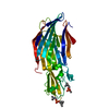
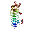
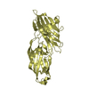
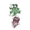
| Title | Crystal structure of red fluorescent protein mCherry complexed with the nanobody LaM4 at 1.9 Angstron resolution | ||||||||||||
 Components Components |
| ||||||||||||
 Keywords Keywords | FLUORESCENT PROTEIN / Complex / Nanobody / red fluorescent protein mCherry / LaM4 | ||||||||||||
| Function / homology |  Function and homology information Function and homology information | ||||||||||||
| Biological species |  Anaplasma marginale (bacteria) Anaplasma marginale (bacteria) | ||||||||||||
| Method |  X-RAY DIFFRACTION / SYNCHROTRON / MOLECULAR REPLACEMENT / Resolution: 1.919 Å X-RAY DIFFRACTION / SYNCHROTRON / MOLECULAR REPLACEMENT / Resolution: 1.919 Å | ||||||||||||
 Authors Authors | Ding, Y. / Wang, Z.Y. / Hu, R.T. / Chen, X. | ||||||||||||
| Funding support |  China, 2items China, 2items
| ||||||||||||
 Citation Citation | Journal: Protein Sci. / Year: 2021 Title: Structural insights into the binding of nanobodies LaM2 and LaM4 to the red fluorescent protein mCherry. Authors: Wang, Z. / Li, L. / Hu, R. / Zhong, P. / Zhang, Y. / Cheng, S. / Jiang, H. / Liu, R. / Ding, Y. | ||||||||||||
| History |
|
- Structure visualization
| Structure viewer | Molecule: MolmilJmol/JSmol |
|---|
- Downloads & links
-Download
| PDBx/mmCIF format | 6ir1.cif.gz | 141.9 KB | Display | PDBx/mmCIF format |
|---|---|---|---|---|
| PDB format | pdb6ir1.ent.gz | 110.3 KB | Display | PDB format |
| PDBx/mmJSON format | 6ir1.json.gz | Tree view | PDBx/mmJSON format | |
| Others |  Other downloads Other downloads |
-Validation report
| Arichive directory | https://data.pdbj.org/pub/pdb/validation_reports/ir/6ir1ftp://data.pdbj.org/pub/pdb/validation_reports/ir/6ir1 | HTTPS FTP |
|---|
-Related structure data
| Related structure data |  6ir2C  2h5qS  3k1kS S: Starting model for refinement C: citing same article ( |
|---|---|
| Similar structure data |


-Links
 PDBj
PDBj


- Assembly
| Deposited unit | 
| ||||||||
|---|---|---|---|---|---|---|---|---|---|
| 1 |
| ||||||||
| Unit cell |
| ||||||||
| Components on special symmetry positions |
|
-Components
| #1: Protein | Mass: 26797.229 Da / Num. of mol.: 1 Source method: isolated from a genetically manipulated source Source: (gene. exp.) Anaplasma marginale (bacteria) / Gene: mCherry / Production host: |
|---|---|
| #2: Antibody | Mass: 14021.442 Da / Num. of mol.: 1 Source method: isolated from a genetically manipulated source Source: (gene. exp.) |
| #3: Water | ChemComp-HOH /  Mass: 18.015 Da / Num. of mol.: 162 / Source method: isolated from a natural source / Formula: H2O Mass: 18.015 Da / Num. of mol.: 162 / Source method: isolated from a natural source / Formula: H2O |
| Has protein modification | Y |
-Experimental details
-Experiment
| Experiment | Method: X-RAY DIFFRACTION / Number of used crystals: 1 |
|---|
- Sample preparation
| Crystal | Density Matthews: 2.14 Å3/Da / Density % sol: 42.42 % |
|---|---|
| Crystal grow | Temperature: 293 K / Method: vapor diffusion, sitting drop / pH: 5.5 Details: 0.2M Lithium sulfate monohydrate, 0.1M BIS-TRIS pH 5.5, 25%(w/v) Polyethylene glycol 3,350. |
-Data collection
| Diffraction | Mean temperature: 100 K / Serial crystal experiment: N | ||||||||||||||||||||||||
|---|---|---|---|---|---|---|---|---|---|---|---|---|---|---|---|---|---|---|---|---|---|---|---|---|---|
| Diffraction source | Source: SYNCHROTRON / Site: SSRF / Beamline: BL17U1 / Wavelength: 0.97917 Å | ||||||||||||||||||||||||
| Detector | Type: DECTRIS EIGER X 16M / Detector: PIXEL / Date: Apr 30, 2018 | ||||||||||||||||||||||||
| Radiation | Protocol: SINGLE WAVELENGTH / Monochromatic (M) / Laue (L): M / Scattering type: x-ray | ||||||||||||||||||||||||
| Radiation wavelength | Wavelength: 0.97917 Å / Relative weight: 1 | ||||||||||||||||||||||||
| Reflection | Resolution: 1.919→40.26 Å / Num. obs: 24985 / % possible obs: 94.1 % / Redundancy: 6.4 % / Biso Wilson estimate: 25.2 Å2 / CC1/2: 0.988 / Rmerge(I) obs: 0.166 / Rpim(I) all: 0.07 / Rrim(I) all: 0.18 / Net I/σ(I): 8 | ||||||||||||||||||||||||
| Reflection shell | Diffraction-ID: 1
|
- Processing
| Software |
| |||||||||||||||||||||||||||||||||||||||||||||||||||||||||||||||
|---|---|---|---|---|---|---|---|---|---|---|---|---|---|---|---|---|---|---|---|---|---|---|---|---|---|---|---|---|---|---|---|---|---|---|---|---|---|---|---|---|---|---|---|---|---|---|---|---|---|---|---|---|---|---|---|---|---|---|---|---|---|---|---|---|
| Refinement | Method to determine structure: MOLECULAR REPLACEMENT Starting model: 2H5Q, 3K1K Resolution: 1.919→32.554 Å / SU ML: 0.24 / Cross valid method: THROUGHOUT / σ(F): 1.35 / Phase error: 26.39
| |||||||||||||||||||||||||||||||||||||||||||||||||||||||||||||||
| Solvent computation | Shrinkage radii: 0.9 Å / VDW probe radii: 1.11 Å | |||||||||||||||||||||||||||||||||||||||||||||||||||||||||||||||
| Displacement parameters | Biso max: 150.2 Å2 / Biso mean: 35.3018 Å2 / Biso min: 9.39 Å2 | |||||||||||||||||||||||||||||||||||||||||||||||||||||||||||||||
| Refinement step | Cycle: final / Resolution: 1.919→32.554 Å
| |||||||||||||||||||||||||||||||||||||||||||||||||||||||||||||||
| Refine LS restraints |
| |||||||||||||||||||||||||||||||||||||||||||||||||||||||||||||||
| LS refinement shell | Refine-ID: X-RAY DIFFRACTION / Rfactor Rfree error: 0 / Total num. of bins used: 8
|
