Mass: 18.015 Da / Num. of mol.: 274 / Source method: isolated from a natural source / Formula: H2O
-
Details
Compound details
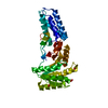
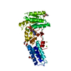
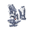
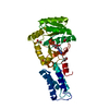
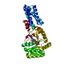
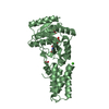
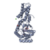
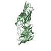
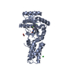
ESSENTIAL FOR EFFICIENT EXPORT OF EXTRA-CYTOPLASMIC PROTEINS. BINDS TO THE SIGNAL SEQUENCE OF ...ESSENTIAL FOR EFFICIENT EXPORT OF EXTRA-CYTOPLASMIC PROTEINS. BINDS TO THE SIGNAL SEQUENCE OF PROTEINS WHEN IT EMERGES FROM THE RIBOSOMES. TWO DOMAIN ARCHITECTURE: NG-DOMAIN BINDS GTP, M-DOMAIN BINDS RNA AND SIGNAL PEPTIDE OF THE PROTEIN TO BE TRANSPORTED. MEMBER OF THE SRP FAMILY OF GTP-BINDING PROTEINS
-
Experimental details
-
Experiment
Experiment
Method: X-RAY DIFFRACTION / Number of used crystals: 1
-
Sample preparation
Crystal
Density Matthews: 2.76 Å3/Da / Density % sol: 55.36 %
Resolution: 2.1→56.8 Å / Cor.coef. Fo:Fc: 0.941 / Cor.coef. Fo:Fc free: 0.913 / SU B: 4.928 / SU ML: 0.133 / Cross valid method: THROUGHOUT / ESU R: 0.22 / ESU R Free: 0.184 / Stereochemistry target values: MAXIMUM LIKELIHOOD / Details: HYDROGENS HAVE BEEN ADDED IN THE RIDING POSITIONS
Rfactor
Num. reflection
% reflection
Selection details
Rfree
0.237
2023
5.1 %
RANDOM
Rwork
0.195
-
-
-
obs
0.197
38000
97.1 %
-
Solvent computation
Ion probe radii: 0.8 Å / Shrinkage radii: 0.8 Å / VDW probe radii: 1.4 Å / Solvent model: BABINET MODEL WITH MASK
 Movie
Movie Controller


 Open data
 Basic information
 Components
 Keywords
 Function and homology information
 THERMUS AQUATICUS (bacteria)
 X-RAY DIFFRACTION /
  Authors
 Citation
 Structure visualization
 Downloads & links
 Other downloads
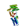


 PDBj
PDBj Assembly


 Mass: 35.453 Da / Num. of mol.: 1 / Source method: obtained synthetically / Formula: Cl
 Mass: 46.025 Da / Num. of mol.: 5 / Source method: obtained synthetically / Formula: CH2O2
 Type: RNA linking / Mass: 443.201 Da / Num. of mol.: 2 / Source method: obtained synthetically / Formula: C10H15N5O11P2 / Comment: GDP, energy-carrying molecule*YM
 Mass: 24.305 Da / Num. of mol.: 4 / Source method: obtained synthetically / Formula: Mg
 Sample preparation
 / Beamline: 5ID-B / Wavelength: 0.96392
  Processing