 Movie
Movie Controller

[English] 日本語
 Yorodumi
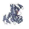
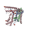
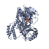
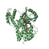
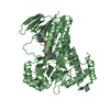
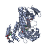
- PDB-4ocl: Crystal Structure of the Rpn8-Rpn11 MPN domain heterodimer, cryst... -
+ Open data

- Basic information
| Entry | Database: PDB / ID: 4ocl | ||||||
|---|---|---|---|---|---|---|---|
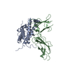
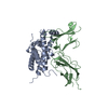
| Title | Crystal Structure of the Rpn8-Rpn11 MPN domain heterodimer, crystal form Ia | ||||||
 Components Components |
| ||||||
 Keywords Keywords | HYDROLASE / PROTEIN BINDING / 26S proteasome / isopeptidase activity / regulatory particle / lid / ubiquitin | ||||||
| Function / homology |  Function and homology information Function and homology information: / proteasome storage granule assembly / peroxisome fission / mitochondrial fission / proteasome regulatory particle, lid subcomplex / Proteasome assembly / Cross-presentation of soluble exogenous antigens (endosomes) / TNFR2 non-canonical NF-kB pathway / metal-dependent deubiquitinase activity / Regulation of PTEN stability and activity ...: / proteasome storage granule assembly / peroxisome fission / mitochondrial fission / proteasome regulatory particle, lid subcomplex / Proteasome assembly / Cross-presentation of soluble exogenous antigens (endosomes) / TNFR2 non-canonical NF-kB pathway / metal-dependent deubiquitinase activity / Regulation of PTEN stability and activity / CDK-mediated phosphorylation and removal of Cdc6 / FBXL7 down-regulates AURKA during mitotic entry and in early mitosis / KEAP1-NFE2L2 pathway / Neddylation / Ubiquitin-Mediated Degradation of Phosphorylated Cdc25A / Orc1 removal from chromatin / MAPK6/MAPK4 signaling / proteasome binding / Antigen processing: Ubiquitination & Proteasome degradation / proteasome storage granule / Ub-specific processing proteases / protein deubiquitination / proteasome assembly / Neutrophil degranulation / proteasome complex / metallopeptidase activity / positive regulation of proteasomal ubiquitin-dependent protein catabolic process / ubiquitin-dependent protein catabolic process / proteasome-mediated ubiquitin-dependent protein catabolic process / cysteine-type deubiquitinase activity / ubiquitinyl hydrolase 1 / mitochondrion / metal ion binding / nucleus / cytosol Similarity search - Function | ||||||
| Biological species |   | ||||||
| Method |  X-RAY DIFFRACTION / SYNCHROTRON / MOLECULAR REPLACEMENT / molecular replacement / Resolution: 2.4 Å X-RAY DIFFRACTION / SYNCHROTRON / MOLECULAR REPLACEMENT / molecular replacement / Resolution: 2.4 Å | ||||||
 Authors Authors | Pathare, G.R. / Bracher, A. | ||||||
 Citation Citation | Journal: Proc.Natl.Acad.Sci.USA / Year: 2014 Title: Crystal structure of the proteasomal deubiquitylation module Rpn8-Rpn11. Authors: Pathare, G.R. / Nagy, I. / Sledz, P. / Anderson, D.J. / Zhou, H.J. / Pardon, E. / Steyaert, J. / Forster, F. / Bracher, A. / Baumeister, W. | ||||||
| History |
|
- Structure visualization
| Structure viewer | Molecule: MolmilJmol/JSmol |
|---|
- Downloads & links
-Download
| PDBx/mmCIF format | 4ocl.cif.gz | 382.8 KB | Display | PDBx/mmCIF format |
|---|---|---|---|---|
| PDB format | pdb4ocl.ent.gz | 311.3 KB | Display | PDB format |
| PDBx/mmJSON format | 4ocl.json.gz | Tree view | PDBx/mmJSON format | |
| Others |  Other downloads Other downloads |
-Validation report
| Arichive directory | https://data.pdbj.org/pub/pdb/validation_reports/oc/4oclftp://data.pdbj.org/pub/pdb/validation_reports/oc/4ocl | HTTPS FTP |
|---|
-Related structure data
| Related structure data | 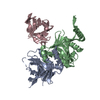 4ocmC 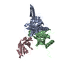 4ocnC 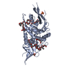 2o95S 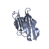 2x1pS C: citing same article ( S: Starting model for refinement |
|---|---|
| Similar structure data |

-Links
 PDBj
PDBj


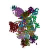
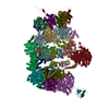
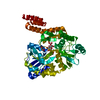
- Assembly
| Deposited unit | 
| ||||||||
|---|---|---|---|---|---|---|---|---|---|
| 1 | 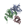
| ||||||||
| 2 | 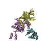
| ||||||||
| Unit cell |
|
-Components
| #1: Protein | Mass: 20838.512 Da / Num. of mol.: 2 / Fragment: UNP residues 1-176 Source method: isolated from a genetically manipulated source Source: (gene. exp.) Strain: ATCC 204508 / S288c / Gene: O5360, RPN8, YOR261C / Plasmid: pRSFDuet / Production host:  #2: Protein | Mass: 24566.283 Da / Num. of mol.: 2 / Fragment: UNP residues 1-220 Source method: isolated from a genetically manipulated source Source: (gene. exp.) Strain: ATCC 204508 / S288c / Gene: MPR1, RPN11, YFR004W / Plasmid: pRSFDuet / Production host: #3: Antibody | Mass: 14854.423 Da / Num. of mol.: 2 Source method: isolated from a genetically manipulated source Source: (gene. exp.) #4: Chemical |   Mass: 65.409 Da / Num. of mol.: 2 / Source method: obtained synthetically / Formula: Zn Mass: 65.409 Da / Num. of mol.: 2 / Source method: obtained synthetically / Formula: Zn#5: Water | ChemComp-HOH / |  Mass: 18.015 Da / Num. of mol.: 94 / Source method: isolated from a natural source / Formula: H2O Mass: 18.015 Da / Num. of mol.: 94 / Source method: isolated from a natural source / Formula: H2OHas protein modification | Y | Sequence details | RPN8 AND RPN11 WERE EXPRESSED AS A FUSION PROTEIN CONNECTED BY A GSGGSGGSG LINKER. THEY HAVE BEEN ...RPN8 AND RPN11 WERE EXPRESSED AS A FUSION PROTEIN CONNECTED BY A GSGGSGGSG LINKER. THEY HAVE BEEN REPRESENTE | |
|---|
-Experimental details
-Experiment
| Experiment | Method: X-RAY DIFFRACTION / Number of used crystals: 1 |
|---|
- Sample preparation
| Crystal | Density Matthews: 2.35 Å3/Da / Density % sol: 47.71 % |
|---|---|
| Crystal grow | Temperature: 291 K / Method: vapor diffusion, sitting drop / pH: 6 Details: 50 mM MES, pH 6.0, 200 mM calcium acetate, 22% PEG3350, VAPOR DIFFUSION, SITTING DROPS, temperature 291K |
-Data collection
| Diffraction | Mean temperature: 100 K | ||||||||||||||||||||||||||||||||||||||||||||||||||||||||||||||||||||||||||||||||||||||||
|---|---|---|---|---|---|---|---|---|---|---|---|---|---|---|---|---|---|---|---|---|---|---|---|---|---|---|---|---|---|---|---|---|---|---|---|---|---|---|---|---|---|---|---|---|---|---|---|---|---|---|---|---|---|---|---|---|---|---|---|---|---|---|---|---|---|---|---|---|---|---|---|---|---|---|---|---|---|---|---|---|---|---|---|---|---|---|---|---|---|
| Diffraction source | Source: SYNCHROTRON / Site: SLS  / Beamline: X10SA / Wavelength: 1.00753 Å / Beamline: X10SA / Wavelength: 1.00753 Å | ||||||||||||||||||||||||||||||||||||||||||||||||||||||||||||||||||||||||||||||||||||||||
| Detector | Type: PSI PILATUS 6M / Detector: PIXEL / Date: May 25, 2013 | ||||||||||||||||||||||||||||||||||||||||||||||||||||||||||||||||||||||||||||||||||||||||
| Radiation | Monochromator: double crystal Si(111) / Protocol: SINGLE WAVELENGTH / Monochromatic (M) / Laue (L): M / Scattering type: x-ray | ||||||||||||||||||||||||||||||||||||||||||||||||||||||||||||||||||||||||||||||||||||||||
| Radiation wavelength | Wavelength: 1.00753 Å / Relative weight: 1 | ||||||||||||||||||||||||||||||||||||||||||||||||||||||||||||||||||||||||||||||||||||||||
| Reflection | Resolution: 2.392→98.44 Å / Num. all: 41691 / Num. obs: 41691 / % possible obs: 96.4 % / Observed criterion σ(F): -100 / Observed criterion σ(I): -100 / Redundancy: 3.5 % / Biso Wilson estimate: 54.346 Å2 / Rmerge(I) obs: 0.081 / Rsym value: 0.081 / Net I/σ(I): 10.1 | ||||||||||||||||||||||||||||||||||||||||||||||||||||||||||||||||||||||||||||||||||||||||
| Reflection shell | Diffraction-ID: 1
|
-Phasing
| Phasing | Method: molecular replacement | |||||||||
|---|---|---|---|---|---|---|---|---|---|---|
| Phasing MR |
|
- Processing
| Software |
| |||||||||||||||||||||||||||||||||||||||||||||||||||||||||||||||||||||||||||||||||||||||||||||||||||||||||||||||||||||||||||||||||||||||||||||||||||||||||||||||||||||||||||||||
|---|---|---|---|---|---|---|---|---|---|---|---|---|---|---|---|---|---|---|---|---|---|---|---|---|---|---|---|---|---|---|---|---|---|---|---|---|---|---|---|---|---|---|---|---|---|---|---|---|---|---|---|---|---|---|---|---|---|---|---|---|---|---|---|---|---|---|---|---|---|---|---|---|---|---|---|---|---|---|---|---|---|---|---|---|---|---|---|---|---|---|---|---|---|---|---|---|---|---|---|---|---|---|---|---|---|---|---|---|---|---|---|---|---|---|---|---|---|---|---|---|---|---|---|---|---|---|---|---|---|---|---|---|---|---|---|---|---|---|---|---|---|---|---|---|---|---|---|---|---|---|---|---|---|---|---|---|---|---|---|---|---|---|---|---|---|---|---|---|---|---|---|---|---|---|---|---|
| Refinement | Method to determine structure: MOLECULAR REPLACEMENT Starting model: PDB ENTRIES 2X1P AND 2O95 Resolution: 2.4→30 Å / Cor.coef. Fo:Fc: 0.948 / Cor.coef. Fo:Fc free: 0.914 / WRfactor Rfree: 0.253 / WRfactor Rwork: 0.1946 / Occupancy max: 1 / Occupancy min: 1 / FOM work R set: 0.8174 / SU B: 19.64 / SU ML: 0.206 / SU R Cruickshank DPI: 0.4107 / SU Rfree: 0.2683 / Cross valid method: THROUGHOUT / σ(F): 0 / ESU R: 0.411 / ESU R Free: 0.268 / Stereochemistry target values: MAXIMUM LIKELIHOOD / Details: HYDROGENS HAVE BEEN ADDED IN THE RIDING POSITIONS
| |||||||||||||||||||||||||||||||||||||||||||||||||||||||||||||||||||||||||||||||||||||||||||||||||||||||||||||||||||||||||||||||||||||||||||||||||||||||||||||||||||||||||||||||
| Solvent computation | Ion probe radii: 0.8 Å / Shrinkage radii: 0.8 Å / VDW probe radii: 1.4 Å / Solvent model: MASK | |||||||||||||||||||||||||||||||||||||||||||||||||||||||||||||||||||||||||||||||||||||||||||||||||||||||||||||||||||||||||||||||||||||||||||||||||||||||||||||||||||||||||||||||
| Displacement parameters | Biso max: 150.67 Å2 / Biso mean: 47.387 Å2 / Biso min: 18.54 Å2
| |||||||||||||||||||||||||||||||||||||||||||||||||||||||||||||||||||||||||||||||||||||||||||||||||||||||||||||||||||||||||||||||||||||||||||||||||||||||||||||||||||||||||||||||
| Refinement step | Cycle: LAST / Resolution: 2.4→30 Å
| |||||||||||||||||||||||||||||||||||||||||||||||||||||||||||||||||||||||||||||||||||||||||||||||||||||||||||||||||||||||||||||||||||||||||||||||||||||||||||||||||||||||||||||||
| Refine LS restraints |
| |||||||||||||||||||||||||||||||||||||||||||||||||||||||||||||||||||||||||||||||||||||||||||||||||||||||||||||||||||||||||||||||||||||||||||||||||||||||||||||||||||||||||||||||
| LS refinement shell | Resolution: 2.4→2.462 Å / Total num. of bins used: 20
| |||||||||||||||||||||||||||||||||||||||||||||||||||||||||||||||||||||||||||||||||||||||||||||||||||||||||||||||||||||||||||||||||||||||||||||||||||||||||||||||||||||||||||||||
| Refinement TLS params. | Method: refined / Refine-ID: X-RAY DIFFRACTION
| |||||||||||||||||||||||||||||||||||||||||||||||||||||||||||||||||||||||||||||||||||||||||||||||||||||||||||||||||||||||||||||||||||||||||||||||||||||||||||||||||||||||||||||||
| Refinement TLS group |
|
