 Movie
Movie Controller

[English] 日本語
 Yorodumi
- PDB-2o95: Crystal Structure of the Metal-Free Dimeric Human Mov34 MPN domai... -
+ Open data

- Basic information
| Entry | Database: PDB / ID: 2o95 | ||||||
|---|---|---|---|---|---|---|---|
| Title | Crystal Structure of the Metal-Free Dimeric Human Mov34 MPN domain (residues 1-186) | ||||||
 Components Components | 26S proteasome non-ATPase regulatory subunit 7 | ||||||
 Keywords Keywords | UNKNOWN FUNCTION / PSMD7 / 26S Proteasome / Mov34 / Jab1/MPN / metal-free dimer | ||||||
| Function / homology |  Function and homology information Function and homology informationproteasome regulatory particle / Regulation of ornithine decarboxylase (ODC) / Proteasome assembly / Cross-presentation of soluble exogenous antigens (endosomes) / Somitogenesis / proteasome complex / Regulation of activated PAK-2p34 by proteasome mediated degradation / Autodegradation of Cdh1 by Cdh1:APC/C / APC/C:Cdc20 mediated degradation of Securin / Asymmetric localization of PCP proteins ...proteasome regulatory particle / Regulation of ornithine decarboxylase (ODC) / Proteasome assembly / Cross-presentation of soluble exogenous antigens (endosomes) / Somitogenesis / proteasome complex / Regulation of activated PAK-2p34 by proteasome mediated degradation / Autodegradation of Cdh1 by Cdh1:APC/C / APC/C:Cdc20 mediated degradation of Securin / Asymmetric localization of PCP proteins / Ubiquitin-dependent degradation of Cyclin D / SCF-beta-TrCP mediated degradation of Emi1 / NIK-->noncanonical NF-kB signaling / AUF1 (hnRNP D0) binds and destabilizes mRNA / TNFR2 non-canonical NF-kB pathway / Assembly of the pre-replicative complex / Vpu mediated degradation of CD4 / Cdc20:Phospho-APC/C mediated degradation of Cyclin A / Dectin-1 mediated noncanonical NF-kB signaling / Degradation of DVL / Degradation of AXIN / Degradation of CRY and PER proteins / Hh mutants are degraded by ERAD / Activation of NF-kappaB in B cells / G2/M Checkpoints / Degradation of GLI1 by the proteasome / Hedgehog ligand biogenesis / Regulation of RUNX3 expression and activity / Autodegradation of the E3 ubiquitin ligase COP1 / Defective CFTR causes cystic fibrosis / Negative regulation of NOTCH4 signaling / GSK3B and BTRC:CUL1-mediated-degradation of NFE2L2 / APC/C:Cdh1 mediated degradation of Cdc20 and other APC/C:Cdh1 targeted proteins in late mitosis/early G1 / Hedgehog 'on' state / Vif-mediated degradation of APOBEC3G / FBXL7 down-regulates AURKA during mitotic entry and in early mitosis / Degradation of GLI2 by the proteasome / GLI3 is processed to GLI3R by the proteasome / Ubiquitin-Mediated Degradation of Phosphorylated Cdc25A / MAPK6/MAPK4 signaling / Degradation of CDH1 / Degradation of beta-catenin by the destruction complex / Oxygen-dependent proline hydroxylation of Hypoxia-inducible Factor Alpha / CDK-mediated phosphorylation and removal of Cdc6 / ABC-family protein mediated transport / CLEC7A (Dectin-1) signaling / SCF(Skp2)-mediated degradation of p27/p21 / FCERI mediated NF-kB activation / Regulation of expression of SLITs and ROBOs / Regulation of PTEN stability and activity / Interleukin-1 signaling / Orc1 removal from chromatin / Regulation of RUNX2 expression and activity / Regulation of RAS by GAPs / The role of GTSE1 in G2/M progression after G2 checkpoint / Separation of Sister Chromatids / UCH proteinases / KEAP1-NFE2L2 pathway / Downstream TCR signaling / Antigen processing: Ubiquitination & Proteasome degradation / RUNX1 regulates transcription of genes involved in differentiation of HSCs / ER-Phagosome pathway / Neddylation / secretory granule lumen / ficolin-1-rich granule lumen / proteasome-mediated ubiquitin-dependent protein catabolic process / Ub-specific processing proteases / Neutrophil degranulation / protein homodimerization activity / extracellular exosome / extracellular region / nucleoplasm / membrane / nucleus / cytosol Similarity search - Function | ||||||
| Biological species |  Homo sapiens (human) Homo sapiens (human) | ||||||
| Method |  X-RAY DIFFRACTION / SYNCHROTRON / SAD / Resolution: 1.95 Å X-RAY DIFFRACTION / SYNCHROTRON / SAD / Resolution: 1.95 Å | ||||||
 Authors Authors | Sanches, M. / Alves, B.S.C. / Zanchin, N.I.T. / Guimaraes, B.G. | ||||||
 Citation Citation | Journal: J.Mol.Biol. / Year: 2007 Title: The Crystal Structure of the Human Mov34 MPN Domain Reveals a Metal-free Dimer Authors: Sanches, M. / Alves, B.S.C. / Zanchin, N.I.T. / Guimaraes, B.G. #1: Journal: Biochem.Biophys.Res.Commun. / Year: 2006 Title: Characterization of the human ortholog of Mov34 reveals eight N-terminal residues important for MPN domain stability Authors: Alves, B.S.C. / Oyama Jr., S. / Gozzo, F.C. / Sanches, M. / Guimaraes, B.G. / Zanchin, N.I.T. | ||||||
| History |
|
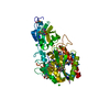
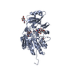
- Structure visualization
| Structure viewer | Molecule: MolmilJmol/JSmol |
|---|
- Downloads & links
-Download
| PDBx/mmCIF format | 2o95.cif.gz | 172.6 KB | Display | PDBx/mmCIF format |
|---|---|---|---|---|
| PDB format | pdb2o95.ent.gz | 135 KB | Display | PDB format |
| PDBx/mmJSON format | 2o95.json.gz | Tree view | PDBx/mmJSON format | |
| Others |  Other downloads Other downloads |
-Validation report
| Arichive directory | https://data.pdbj.org/pub/pdb/validation_reports/o9/2o95ftp://data.pdbj.org/pub/pdb/validation_reports/o9/2o95 | HTTPS FTP |
|---|
-Related structure data


-Links
 PDBj
PDBj


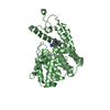
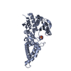
- Assembly
| Deposited unit | 
| ||||||||||||||||||
|---|---|---|---|---|---|---|---|---|---|---|---|---|---|---|---|---|---|---|---|
| 1 | 
| ||||||||||||||||||
| 2 | 
| ||||||||||||||||||
| 3 |
| ||||||||||||||||||
| 4 | 
| ||||||||||||||||||
| Unit cell |
| ||||||||||||||||||
| Noncrystallographic symmetry (NCS) | NCS domain:
NCS domain segments: Component-ID: 1 / Ens-ID: 1 / Beg auth comp-ID: MET / Beg label comp-ID: MET / End auth comp-ID: ASP / End label comp-ID: ASP / Refine code: 1 / Auth seq-ID: 1 - 181 / Label seq-ID: 2 - 182
| ||||||||||||||||||
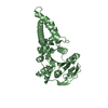
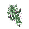
| Details | The biological assembly is a dimer generated by the symmetry operation -x y -z. The asymmetric unit contains a dimmer which does not represent the biological assembly. |
-Components
-Protein , 1 types, 2 molecules AB
| #1: Protein | Mass: 21080.977 Da / Num. of mol.: 2 / Fragment: MPN domain, N-terminus domain, residues 1-186 Source method: isolated from a genetically manipulated source Source: (gene. exp.) Homo sapiens (human) / Gene: PSMD7, Mov34L / Plasmid: pET28a-TEV / Production host:  |
|---|
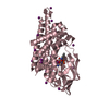
-Non-polymers , 6 types, 330 molecules 


| #2: Chemical |  Mass: 96.063 Da / Num. of mol.: 3 / Source method: obtained synthetically / Formula: SO4 Mass: 96.063 Da / Num. of mol.: 3 / Source method: obtained synthetically / Formula: SO4#3: Chemical | ChemComp-12P / |  Mass: 546.646 Da / Num. of mol.: 1 / Source method: obtained synthetically / Formula: C24H50O13 / Comment: precipitant*YM Mass: 546.646 Da / Num. of mol.: 1 / Source method: obtained synthetically / Formula: C24H50O13 / Comment: precipitant*YM#4: Chemical | ChemComp-PG4 / |  Mass: 194.226 Da / Num. of mol.: 1 / Source method: obtained synthetically / Formula: C8H18O5 / Comment: precipitant*YM Mass: 194.226 Da / Num. of mol.: 1 / Source method: obtained synthetically / Formula: C8H18O5 / Comment: precipitant*YM#5: Chemical | ChemComp-PGE /  Mass: 150.173 Da / Num. of mol.: 4 / Source method: obtained synthetically / Formula: C6H14O4 Mass: 150.173 Da / Num. of mol.: 4 / Source method: obtained synthetically / Formula: C6H14O4#6: Chemical | ChemComp-ETE / |  Mass: 208.252 Da / Num. of mol.: 1 / Source method: obtained synthetically / Formula: C9H20O5 Mass: 208.252 Da / Num. of mol.: 1 / Source method: obtained synthetically / Formula: C9H20O5#7: Water | ChemComp-HOH / | Mass: 18.015 Da / Num. of mol.: 320 / Source method: isolated from a natural source / Formula: H2O |
|---|
-Experimental details
-Experiment
| Experiment | Method: X-RAY DIFFRACTION / Number of used crystals: 1 |
|---|
- Sample preparation
| Crystal | Density Matthews: 3.25 Å3/Da / Density % sol: 62.2 % |
|---|---|
| Crystal grow | Temperature: 291 K / Method: vapor diffusion, hanging drop / pH: 4.2 Details: 0.1M phosphate-citrate buffer, 20% PEG 1000, 0.2M LiSO4, 10mM MnCl2 as additive, pH 4.2, VAPOR DIFFUSION, HANGING DROP, temperature 291K |
-Data collection
| Diffraction |
| |||||||||
|---|---|---|---|---|---|---|---|---|---|---|
| Diffraction source | Source: SYNCHROTRON / Site: LNLS  / Beamline: D03B-MX1 / Wavelength: 1.427 Å / Beamline: D03B-MX1 / Wavelength: 1.427 Å | |||||||||
| Detector | Type: MAR CCD 165 mm / Detector: CCD / Date: Jun 6, 2006 | |||||||||
| Radiation | Monochromator: Si 111 CHANNEL / Protocol: SINGLE WAVELENGTH / Monochromatic (M) / Laue (L): M / Scattering type: x-ray | |||||||||
| Radiation wavelength | Wavelength: 1.427 Å / Relative weight: 1 | |||||||||
| Reflection | Resolution: 1.95→68.205 Å / Num. all: 49805 / Num. obs: 39272 / % possible obs: 99.7 % / Observed criterion σ(F): 0 / Observed criterion σ(I): 1.6 / Redundancy: 3.3 % / Biso Wilson estimate: 34.2 Å2 / Rmerge(I) obs: 0.05 / Rsym value: 0.05 / Net I/σ(I): 11.9 | |||||||||
| Reflection shell | Resolution: 1.95→2.06 Å / Redundancy: 3.1 % / Rmerge(I) obs: 0.476 / Mean I/σ(I) obs: 1.6 / Num. measured all: 17623 / Num. unique all: 5682 / Rsym value: 0.476 / % possible all: 99.5 |
-Phasing
| Phasing | Method: SAD | ||||||||||||||||||||||||||||||||||||||||||||||||||||||||||||||||||||||||||||||||||||||||||||||||||||||||||||||||||||||||||||||||||||||||||||||||||||||||||||||||||||||||||||||||||||||||||||||||||||||||||||||||||||||||||||||||||||||||||||||||||||||
|---|---|---|---|---|---|---|---|---|---|---|---|---|---|---|---|---|---|---|---|---|---|---|---|---|---|---|---|---|---|---|---|---|---|---|---|---|---|---|---|---|---|---|---|---|---|---|---|---|---|---|---|---|---|---|---|---|---|---|---|---|---|---|---|---|---|---|---|---|---|---|---|---|---|---|---|---|---|---|---|---|---|---|---|---|---|---|---|---|---|---|---|---|---|---|---|---|---|---|---|---|---|---|---|---|---|---|---|---|---|---|---|---|---|---|---|---|---|---|---|---|---|---|---|---|---|---|---|---|---|---|---|---|---|---|---|---|---|---|---|---|---|---|---|---|---|---|---|---|---|---|---|---|---|---|---|---|---|---|---|---|---|---|---|---|---|---|---|---|---|---|---|---|---|---|---|---|---|---|---|---|---|---|---|---|---|---|---|---|---|---|---|---|---|---|---|---|---|---|---|---|---|---|---|---|---|---|---|---|---|---|---|---|---|---|---|---|---|---|---|---|---|---|---|---|---|---|---|---|---|---|---|---|---|---|---|---|---|---|---|---|---|---|---|---|---|---|---|
| Phasing MAD set |
| ||||||||||||||||||||||||||||||||||||||||||||||||||||||||||||||||||||||||||||||||||||||||||||||||||||||||||||||||||||||||||||||||||||||||||||||||||||||||||||||||||||||||||||||||||||||||||||||||||||||||||||||||||||||||||||||||||||||||||||||||||||||
| Phasing MAD set shell |
| ||||||||||||||||||||||||||||||||||||||||||||||||||||||||||||||||||||||||||||||||||||||||||||||||||||||||||||||||||||||||||||||||||||||||||||||||||||||||||||||||||||||||||||||||||||||||||||||||||||||||||||||||||||||||||||||||||||||||||||||||||||||
| Phasing MAD set site |
|
- Processing
| Software |
| ||||||||||||||||||||||||||||||||||||||||||||||||||||||||||||||||||||||||||||||||||||||||||
|---|---|---|---|---|---|---|---|---|---|---|---|---|---|---|---|---|---|---|---|---|---|---|---|---|---|---|---|---|---|---|---|---|---|---|---|---|---|---|---|---|---|---|---|---|---|---|---|---|---|---|---|---|---|---|---|---|---|---|---|---|---|---|---|---|---|---|---|---|---|---|---|---|---|---|---|---|---|---|---|---|---|---|---|---|---|---|---|---|---|---|---|
| Refinement | Method to determine structure: SAD / Resolution: 1.95→30.21 Å / Cor.coef. Fo:Fc: 0.947 / Cor.coef. Fo:Fc free: 0.926 / SU B: 6.712 / SU ML: 0.101 / Isotropic thermal model: ISOTROPIC / Cross valid method: THROUGHOUT / σ(F): 0 / σ(I): 0 / ESU R: 0.148 / ESU R Free: 0.142 / Stereochemistry target values: MAXIMUM LIKELIHOOD / Details: HYDROGENS HAVE BEEN ADDED IN THE RIDING POSITIONS
| ||||||||||||||||||||||||||||||||||||||||||||||||||||||||||||||||||||||||||||||||||||||||||
| Solvent computation | Ion probe radii: 0.8 Å / Shrinkage radii: 0.8 Å / VDW probe radii: 1.4 Å / Solvent model: MASK | ||||||||||||||||||||||||||||||||||||||||||||||||||||||||||||||||||||||||||||||||||||||||||
| Displacement parameters | Biso mean: 27.953 Å2
| ||||||||||||||||||||||||||||||||||||||||||||||||||||||||||||||||||||||||||||||||||||||||||
| Refinement step | Cycle: LAST / Resolution: 1.95→30.21 Å
| ||||||||||||||||||||||||||||||||||||||||||||||||||||||||||||||||||||||||||||||||||||||||||
| Refine LS restraints |
| ||||||||||||||||||||||||||||||||||||||||||||||||||||||||||||||||||||||||||||||||||||||||||
| Refine LS restraints NCS | Dom-ID: 1 / Auth asym-ID: A / Ens-ID: 1 / Number: 1362 / Refine-ID: X-RAY DIFFRACTION
| ||||||||||||||||||||||||||||||||||||||||||||||||||||||||||||||||||||||||||||||||||||||||||
| LS refinement shell | Resolution: 1.95→2.001 Å / Total num. of bins used: 20
|
