 Movie
Movie Controller

[English] 日本語
 Yorodumi
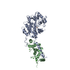
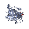
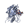
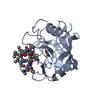
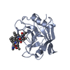
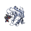
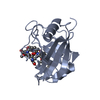
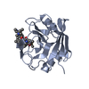
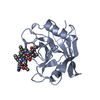
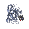
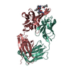
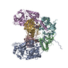
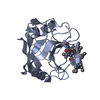
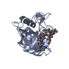
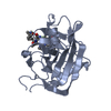
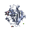
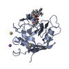
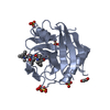
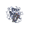
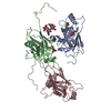
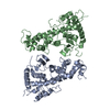
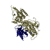
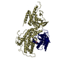
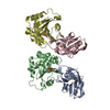
- PDB-1mf8: Crystal Structure of human calcineurin complexed with cyclosporin... -
+ Open data

- Basic information
| Entry | Database: PDB / ID: 1mf8 | ||||||
|---|---|---|---|---|---|---|---|
| Title | Crystal Structure of human calcineurin complexed with cyclosporin A and human cyclophilin | ||||||
 Components Components |
| ||||||
 Keywords Keywords | HYDROLASE/ISOMERASE/IMMUNOSUPPRESSANT / HYDROLASE-ISOMERASE-IMMUNOSUPPRESSANT COMPLEX / CALCINEURIN-CYCLOPHILIN-CYCLOSPORIN COMPLEX / CYCLOSPORIN A / IMMUNOSUPPRESSANT / HYDROLASE / ISOMERASE | ||||||
| Function / homology |  Function and homology information Function and homology informationnegative regulation of angiotensin-activated signaling pathway / calcium-dependent protein serine/threonine phosphatase regulator activity / regulation of cell proliferation involved in kidney morphogenesis / positive regulation of glomerulus development / negative regulation of calcium ion import across plasma membrane / protein serine/threonine phosphatase complex / negative regulation of signaling / calcium-dependent protein serine/threonine phosphatase activity / positive regulation of saliva secretion / peptidyl-serine dephosphorylation ...negative regulation of angiotensin-activated signaling pathway / calcium-dependent protein serine/threonine phosphatase regulator activity / regulation of cell proliferation involved in kidney morphogenesis / positive regulation of glomerulus development / negative regulation of calcium ion import across plasma membrane / protein serine/threonine phosphatase complex / negative regulation of signaling / calcium-dependent protein serine/threonine phosphatase activity / positive regulation of saliva secretion / peptidyl-serine dephosphorylation / calmodulin-dependent protein phosphatase activity / calcineurin complex / positive regulation of connective tissue replacement / positive regulation of calcium ion-dependent exocytosis of neurotransmitter / positive regulation of calcium ion import across plasma membrane / positive regulation of cardiac muscle hypertrophy in response to stress / lung epithelial cell differentiation / negative regulation of dendrite morphogenesis / renal filtration / calcineurin-NFAT signaling cascade / positive regulation of calcineurin-NFAT signaling cascade / negative regulation of protein K48-linked ubiquitination / regulation of apoptotic signaling pathway / cell adhesion molecule production / lipid droplet organization / myelination in peripheral nervous system / skeletal muscle tissue regeneration / transition between fast and slow fiber / negative regulation of viral life cycle / heparan sulfate binding / regulation of viral genome replication / dephosphorylation / positive regulation of osteoclast differentiation / cardiac muscle hypertrophy in response to stress / virion binding / leukocyte chemotaxis / negative regulation of stress-activated MAPK cascade / activation of protein kinase B activity / regulation of synaptic vesicle cycle / endothelial cell activation / positive regulation of activated T cell proliferation / protein dephosphorylation / Basigin interactions / extrinsic component of plasma membrane / protein peptidyl-prolyl isomerization / cyclosporin A binding / Minus-strand DNA synthesis / Plus-strand DNA synthesis / branching involved in blood vessel morphogenesis / dendrite morphogenesis / Uncoating of the HIV Virion / protein-serine/threonine phosphatase / regulation of postsynaptic neurotransmitter receptor internalization / CLEC7A (Dectin-1) induces NFAT activation / Early Phase of HIV Life Cycle / Integration of provirus / APOBEC3G mediated resistance to HIV-1 infection / negative regulation of protein phosphorylation / protein serine/threonine phosphatase activity / parallel fiber to Purkinje cell synapse / calcineurin-mediated signaling / epithelial to mesenchymal transition / viral release from host cell / epidermis development / Binding and entry of HIV virion / Calcineurin activates NFAT / Activation of BAD and translocation to mitochondria / DARPP-32 events / positive regulation of osteoblast differentiation / positive regulation of endocytosis / multicellular organismal response to stress / negative regulation of oxidative stress-induced intrinsic apoptotic signaling pathway / negative regulation of protein kinase activity / postsynaptic modulation of chemical synaptic transmission / phosphatase binding / positive regulation of viral genome replication / keratinocyte differentiation / skeletal muscle fiber development / neutrophil chemotaxis / FCERI mediated Ca+2 mobilization / Gene and protein expression by JAK-STAT signaling after Interleukin-12 stimulation / positive regulation of cell adhesion / : / T cell activation / hippocampal mossy fiber to CA3 synapse / peptidylprolyl isomerase / positive regulation of protein secretion / peptidyl-prolyl cis-trans isomerase activity / wound healing / excitatory postsynaptic potential / G1/S transition of mitotic cell cycle / Assembly Of The HIV Virion / platelet activation / Budding and maturation of HIV virion / sarcolemma / positive regulation of protein phosphorylation / response to calcium ion / platelet aggregation / integrin binding / modulation of chemical synaptic transmission Similarity search - Function | ||||||
| Biological species |  HOMO SAPIENS (human) HOMO SAPIENS (human) TOLYPOCLADIUM INFLATUM (fungus) TOLYPOCLADIUM INFLATUM (fungus) | ||||||
| Method |  X-RAY DIFFRACTION / SYNCHROTRON / MOLECULAR REPLACEMENT / Resolution: 3.1 Å X-RAY DIFFRACTION / SYNCHROTRON / MOLECULAR REPLACEMENT / Resolution: 3.1 Å | ||||||
 Authors Authors | Jin, L. / Harrison, S.C. | ||||||
 Citation Citation | Journal: Proc.Natl.Acad.Sci.USA / Year: 2002 Title: Crystal Structure of Human Calcineurin Complexed with Cyclosporin a and Human Cyclophilin Authors: Jin, L. / Harrison, S.C. | ||||||
| History |
|
- Structure visualization
| Structure viewer | Molecule: MolmilJmol/JSmol |
|---|
- Downloads & links
-Download
| PDBx/mmCIF format | 1mf8.cif.gz | 154.4 KB | Display | PDBx/mmCIF format |
|---|---|---|---|---|
| PDB format | pdb1mf8.ent.gz | 118.1 KB | Display | PDB format |
| PDBx/mmJSON format | 1mf8.json.gz | Tree view | PDBx/mmJSON format | |
| Others |  Other downloads Other downloads |
-Validation report
| Arichive directory | https://data.pdbj.org/pub/pdb/validation_reports/mf/1mf8ftp://data.pdbj.org/pub/pdb/validation_reports/mf/1mf8 | HTTPS FTP |
|---|
-Related structure data


-Links
 PDBj
PDBj


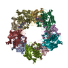
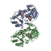
- Assembly
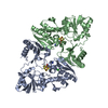
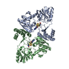
| Deposited unit | 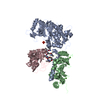
| ||||||||
|---|---|---|---|---|---|---|---|---|---|
| 1 |
| ||||||||
| Unit cell |
|
-Components
-Protein , 3 types, 3 molecules ABC
| #1: Protein | Mass: 42770.637 Da / Num. of mol.: 1 / Fragment: TRUNCATED FORM (RESIDUES 20-392) Source method: isolated from a genetically manipulated source Source: (gene. exp.) HOMO SAPIENS (human) / Plasmid: PETCN ALPHA / Production host:  References: UniProt: Q08209, protein-serine/threonine phosphatase |
|---|---|
| #2: Protein | Mass: 19322.904 Da / Num. of mol.: 1 Source method: isolated from a genetically manipulated source Source: (gene. exp.) HOMO SAPIENS (human) / Plasmid: PETCN ALPHA / Production host: References: UniProt: P63098, protein-serine/threonine phosphatase |
| #3: Protein | Mass: 18036.504 Da / Num. of mol.: 1 Source method: isolated from a genetically manipulated source Source: (gene. exp.) HOMO SAPIENS (human) / Plasmid: PET3A.CYPA / Production host: |
-Protein/peptide , 1 types, 1 molecules D
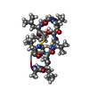
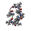
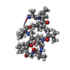
| #4: Protein/peptide | 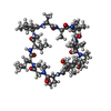  Type: Cyclic peptide / Class: Immunosuppressant / Mass: 1220.625 Da / Num. of mol.: 1 / Source method: obtained synthetically Type: Cyclic peptide / Class: Immunosuppressant / Mass: 1220.625 Da / Num. of mol.: 1 / Source method: obtained syntheticallyDetails: CYCLOSPORIN IS A CYCLIC UNDECAPEPTIDE. CYCLIZATION IS ACHIEVED BY LINKING THE N- AND THE C- TERMINI. Source: (synth.) TOLYPOCLADIUM INFLATUM (fungus) / References: NOR: NOR00033, Cyclosporin A |
|---|
-Non-polymers , 2 types, 5 molecules 

| #5: Chemical | ChemComp-PO4 /  Mass: 94.971 Da / Num. of mol.: 1 / Source method: obtained synthetically / Formula: PO4 Mass: 94.971 Da / Num. of mol.: 1 / Source method: obtained synthetically / Formula: PO4 |
|---|---|
| #6: Chemical | ChemComp-CA /  Mass: 40.078 Da / Num. of mol.: 4 / Source method: obtained synthetically / Formula: Ca Mass: 40.078 Da / Num. of mol.: 4 / Source method: obtained synthetically / Formula: Ca |
-Details
| Compound details | CYCLOSPORI| Has protein modification | Y | |
|---|
-Experimental details
-Experiment
| Experiment | Method: X-RAY DIFFRACTION / Number of used crystals: 1 |
|---|
- Sample preparation
| Crystal | Density Matthews: 2.6 Å3/Da / Density % sol: 46 % | ||||||||||||||||||||||||||||||||||||||||||
|---|---|---|---|---|---|---|---|---|---|---|---|---|---|---|---|---|---|---|---|---|---|---|---|---|---|---|---|---|---|---|---|---|---|---|---|---|---|---|---|---|---|---|---|
| Crystal grow | pH: 4.6 Details: 12% PEG 4000, 5MM POTASSIUM PHOSPHATE, 75MM SODIUM CITRATE, 15% GLYCEROL, 5MM TRIS, PH 4.6, MICROBATCH, TEMPERATURE 292K | ||||||||||||||||||||||||||||||||||||||||||
| Crystal grow | *PLUS Temperature: 19 ℃ / Method: batch method | ||||||||||||||||||||||||||||||||||||||||||
| Components of the solutions | *PLUS
|
-Data collection
| Diffraction | Mean temperature: 100 K |
|---|---|
| Diffraction source | Source: SYNCHROTRON / Site: CHESS  / Beamline: F1 / Wavelength: 0.91611 / Beamline: F1 / Wavelength: 0.91611 |
| Detector | Type: ADSC QUANTUM 4 / Detector: CCD / Date: Mar 12, 2002 |
| Radiation | Monochromator: SI 111 CHANNEL / Protocol: SINGLE WAVELENGTH / Monochromatic (M) / Laue (L): M / Scattering type: x-ray |
| Radiation wavelength | Wavelength: 0.91611 Å / Relative weight: 1 |
| Reflection | Resolution: 3.1→15 Å / Num. obs: 14195 / % possible obs: 95.6 % / Observed criterion σ(I): 0 / Redundancy: 5.3 % / Rsym value: 0.077 / Net I/σ(I): 19.3 |
| Reflection shell | Resolution: 3.1→3.21 Å / Mean I/σ(I) obs: 3.3 / Rsym value: 0.37 / % possible all: 81.7 |
| Reflection | *PLUS Lowest resolution: 15 Å / Rmerge(I) obs: 0.077 |
| Reflection shell | *PLUS Highest resolution: 3.1 Å / % possible obs: 81.7 % / Rmerge(I) obs: 0.377 |
- Processing
| Software |
| ||||||||||||||||||||||||||||||||||||||||||||||||||||||||||||
|---|---|---|---|---|---|---|---|---|---|---|---|---|---|---|---|---|---|---|---|---|---|---|---|---|---|---|---|---|---|---|---|---|---|---|---|---|---|---|---|---|---|---|---|---|---|---|---|---|---|---|---|---|---|---|---|---|---|---|---|---|---|
| Refinement | Method to determine structure: MOLECULAR REPLACEMENT Starting model: PDB ENTRY 1AUI AND 2RMA Resolution: 3.1→15 Å / Cross valid method: THROUGHOUT / σ(F): 0 / Stereochemistry target values: ENGH & HUBER
| ||||||||||||||||||||||||||||||||||||||||||||||||||||||||||||
| Refinement step | Cycle: LAST / Resolution: 3.1→15 Å
| ||||||||||||||||||||||||||||||||||||||||||||||||||||||||||||
| Refine LS restraints |
| ||||||||||||||||||||||||||||||||||||||||||||||||||||||||||||
| Refinement | *PLUS Lowest resolution: 15 Å / Rfactor obs: 0.26 / Rfactor Rfree: 0.3 / Rfactor Rwork: 0.255 | ||||||||||||||||||||||||||||||||||||||||||||||||||||||||||||
| Solvent computation | *PLUS | ||||||||||||||||||||||||||||||||||||||||||||||||||||||||||||
| Displacement parameters | *PLUS | ||||||||||||||||||||||||||||||||||||||||||||||||||||||||||||
| Refine LS restraints | *PLUS Type: c_bond_d / Dev ideal: 0.01 | ||||||||||||||||||||||||||||||||||||||||||||||||||||||||||||
| LS refinement shell | *PLUS Highest resolution: 3.1 Å / Lowest resolution: 3.21 Å / Rfactor Rfree: 0.45 / Rfactor Rwork: 0.409 |
