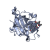
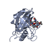
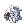
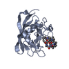
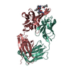
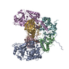
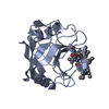
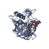
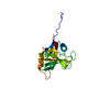
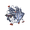
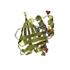
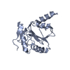
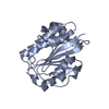
Mass: 18257.805 Da / Num. of mol.: 1 Source method: isolated from a genetically manipulated source Source: (gene. exp.) HOMO SAPIENS (human) / Plasmid: PGEX-6-P1-PPIL1 / Production host: ESCHERICHIA COLI (E. coli) / Strain (production host): ROSETTA2 / Variant (production host): DE3 / References: UniProt: Q9Y3C6, peptidylprolyl isomerase
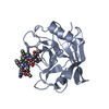
#2: Protein/peptide
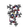
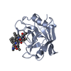
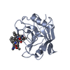
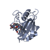
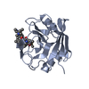
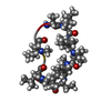
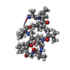
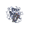
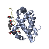
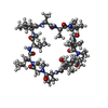
CYCLOSPORINA / CICLOSPORIN / CICLOSPORINE
Type: Cyclic peptide / Class: Immunosuppressant / Mass: 1220.625 Da / Num. of mol.: 1 / Source method: obtained synthetically Details: CYCLOSPORIN IS A CYCLIC UNDECAPEPTIDE. CYCLIZATION IS ACHIEVED BY LINKING THE N- AND THE C- TERMINI. Source: (synth.) TOLYPOCLADIUM INFLATUM (fungus) / References: NOR: NOR00033, Cyclosporin A
Mass: 18.015 Da / Num. of mol.: 294 / Source method: isolated from a natural source / Formula: H2O
Compound details
CYCLOSPORIN IS A CYCLIC UNDECAPEPTIDE. HERE, CYCLOSPORIN A IS REPRESENTED BY THE SEQUENCE (SEQRES) ...CYCLOSPORIN IS A CYCLIC UNDECAPEPTIDE. HERE, CYCLOSPORIN A IS REPRESENTED BY THE SEQUENCE (SEQRES) GROUP: 1 NAME: CYCLOSPORIN A CHAIN: B COMPONENT_1: PEPTIDE LIKE SEQUENCE RESIDUES 1 TO 11 DESCRIPTION: CYCLOSPORIN IS A CYCLIC UNDECAPEPTIDE. CYCLIZATION IS ACHIEVED BY LINKING THE N- AND THE C- TERMINI.
Has protein modification
Y
-
Experimental details
-
Experiment
Experiment
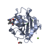
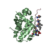
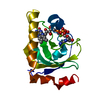
Method: X-RAY DIFFRACTION / Number of used crystals: 1
-
Sample preparation
Crystal
Density Matthews: 2.32 Å3/Da / Density % sol: 47 % / Description: NONE
Crystal grow
Temperature: 277 K / Method: vapor diffusion, hanging drop / pH: 7.2 Details: 0.1 M HEPES, PH 7.2, 0.7 M SODIUM ACETATE, 15 MM CDCL2 AND 50 MM GUANIDINE-HCL GROWN AT 4DEGC
In the structure databanks used in Yorodumi, some data are registered as the other names, "COVID-19 virus" and "2019-nCoV". Here are the details of the virus and the list of structure data.
Jan 31, 2019. EMDB accession codes are about to change! (news from PDBe EMDB page)
EMDB accession codes are about to change! (news from PDBe EMDB page)
The allocation of 4 digits for EMDB accession codes will soon come to an end. Whilst these codes will remain in use, new EMDB accession codes will include an additional digit and will expand incrementally as the available range of codes is exhausted. The current 4-digit format prefixed with “EMD-” (i.e. EMD-XXXX) will advance to a 5-digit format (i.e. EMD-XXXXX), and so on. It is currently estimated that the 4-digit codes will be depleted around Spring 2019, at which point the 5-digit format will come into force.
The EM Navigator/Yorodumi systems omit the EMD- prefix.
Related info.:Q: What is EMD? / ID/Accession-code notation in Yorodumi/EM Navigator
Yorodumi is a browser for structure data from EMDB, PDB, SASBDB, etc.
This page is also the successor to EM Navigator detail page, and also detail information page/front-end page for Omokage search.
The word "yorodu" (or yorozu) is an old Japanese word meaning "ten thousand". "mi" (miru) is to see.
Related info.:EMDB / PDB / SASBDB / Comparison of 3 databanks / Yorodumi Search / Aug 31, 2016. New EM Navigator & Yorodumi / Yorodumi Papers / Jmol/JSmol / Function and homology information / Changes in new EM Navigator and Yorodumi
 Movie
Movie Controller

 Yorodumi
 Open data
 Basic information
 Components
 Keywords
 Function and homology information
 HOMO SAPIENS (human)
 TOLYPOCLADIUM INFLATUM (fungus)
 X-RAY DIFFRACTION /
  Authors
 Citation
 Structure visualization
 Downloads & links
 Other downloads


 PDBj
PDBj


 Assembly
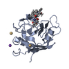

 Type: Cyclic peptide / Class: Immunosuppressant / Mass: 1220.625 Da / Num. of mol.: 1 / Source method: obtained synthetically
 Mass: 112.411 Da / Num. of mol.: 2 / Source method: obtained synthetically / Formula: Cd
 Mass: 22.990 Da / Num. of mol.: 1 / Source method: obtained synthetically / Formula: Na
 Mass: 18.015 Da / Num. of mol.: 294 / Source method: isolated from a natural source / Formula: H2O
 Sample preparation
 / Beamline: 14.2 / Wavelength: 0.9
  Processing