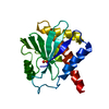
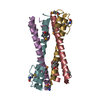
Mass: 15080.600 Da / Num. of mol.: 2 Source method: isolated from a genetically manipulated source Source: (gene. exp.) Lama glama (llama) / Production host: Escherichia coli (E. coli)
#2: Protein
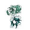
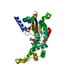
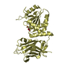
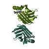
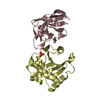
Ricin
Mass: 21538.275 Da / Num. of mol.: 2 Source method: isolated from a genetically manipulated source Source: (gene. exp.) Ricinus communis (castor bean) / Production host: Escherichia coli (E. coli) / References: UniProt: P02879, rRNA N-glycosylase
Resolution: 2.73→65.22 Å / Cor.coef. Fo:Fc: 0.907 / Cor.coef. Fo:Fc free: 0.868 / Cross valid method: THROUGHOUT / ESU R Free: 0.417 / Details: HYDROGENS HAVE BEEN ADDED IN THE RIDING POSITIONS
Rfactor
Num. reflection
% reflection
Selection details
Rfree
0.2667
783
5.1 %
RANDOM
Rwork
0.22634
-
-
-
obs
0.22838
14603
99.65 %
-
Solvent computation
Ion probe radii: 0.8 Å / Shrinkage radii: 0.8 Å / VDW probe radii: 1.2 Å
 Movie
Movie Controller


 Open data
 Basic information
 Components
 Keywords
 Function and homology information
 Ricinus communis (castor bean)
 X-RAY DIFFRACTION /
  Authors
 United States, 1items
  Citation
 Structure visualization
 Downloads & links
 Other downloads

 PDBj
PDBj


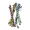
 Assembly


 Mass: 96.063 Da / Num. of mol.: 2 / Source method: obtained synthetically / Formula: SO4
 Mass: 18.015 Da / Num. of mol.: 78 / Source method: isolated from a natural source / Formula: H2O
 Sample preparation
 Processing