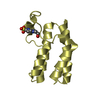
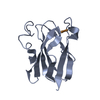
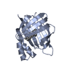
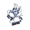
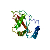
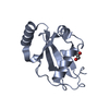
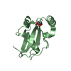
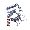
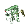
Microtubule-associatedproteins1A/1Blightchain3B / Autophagy-related protein LC3 B / Autophagy-related ubiquitin-like modifier LC3 B / MAP1 light ...Autophagy-related protein LC3 B / Autophagy-related ubiquitin-like modifier LC3 B / MAP1 light chain 3-like protein 2 / MAP1A/MAP1B light chain 3 B / MAP1A/MAP1B LC3 B / Microtubule-associated protein 1 light chain 3 beta
Mass: 14637.859 Da / Num. of mol.: 2 Source method: isolated from a genetically manipulated source Source: (gene. exp.) Mus musculus (house mouse) / Gene: Map1lc3b, Map1alc3, Map1lc3 / Production host: Escherichia coli (E. coli) / References: UniProt: Q9CQV6
Protocol: SINGLE WAVELENGTH / Monochromatic (M) / Laue (L): M / Scattering type: x-ray
Radiation wavelength
Wavelength: 1 Å / Relative weight: 1
Reflection
Resolution: 1.9→50 Å / Num. obs: 21371 / % possible obs: 98.1 % / Redundancy: 3.4 % / Net I/σ(I): 20.2
-
Processing
Software
Name
Version
Classification
REFMAC
5.8.0049
refinement
DENZO
datareduction
SCALEPACK
datascaling
MOLREP
phasing
Refinement
Resolution: 1.9→50 Å / Cor.coef. Fo:Fc: 0.961 / Cor.coef. Fo:Fc free: 0.934 / SU B: 3.974 / SU ML: 0.116 / Cross valid method: THROUGHOUT / ESU R: 0.168 / ESU R Free: 0.161 / Details: HYDROGENS HAVE BEEN ADDED IN THE RIDING POSITIONS
Rfactor
Num. reflection
% reflection
Selection details
Rfree
0.24418
1087
5.1 %
RANDOM
Rwork
0.1881
-
-
-
obs
0.19088
20284
97.99 %
-
Solvent computation
Ion probe radii: 0.8 Å / Shrinkage radii: 0.8 Å / VDW probe radii: 1.2 Å
 Movie
Movie Controller


 Open data
 Basic information
 Components
 Keywords
 Function and homology information
 X-RAY DIFFRACTION /
  Authors
 Citation
 Structure visualization
 Downloads & links
 Other downloads
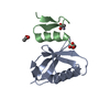

 PDBj
PDBj

 Assembly
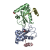


 Mass: 92.094 Da / Num. of mol.: 2 / Source method: obtained synthetically / Formula: C3H8O3
 Mass: 18.015 Da / Num. of mol.: 50 / Source method: isolated from a natural source / Formula: H2O
 Sample preparation
 / Beamline: BL41XU / Wavelength: 1 Å
 Processing