- PDB-4xkl: Crystal structure of NDP52 ZF2 in complex with mono-ubiquitin -
+
Open data
ID or keywords:
Loading...
-
Basic information
Entry
Database: PDB / ID: 4xkl
Title
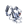
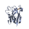
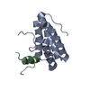
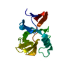
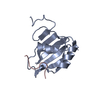
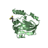
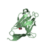
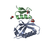
Crystal structure of NDP52 ZF2 in complex with mono-ubiquitin
Components
Calcium-binding and coiled-coil domain-containing protein 2
Ubiquitin
Keywords
PROTEIN BINDING/METAL BINDING PROTEIN / NDP52 / ubiquitin / zinc finger / autophagy receptor / complex / PROTEIN BINDING-METAL BINDING PROTEIN complex
Function / homology
Function and homology information
xenophagy / response to insecticide / positive regulation of autophagosome maturation / autophagosome membrane / viral process / Peptide chain elongation / Selenocysteine synthesis / Formation of a pool of free 40S subunits / Eukaryotic Translation Termination / response to type II interferon ...xenophagy / response to insecticide / positive regulation of autophagosome maturation / autophagosome membrane / viral process / Peptide chain elongation / Selenocysteine synthesis / Formation of a pool of free 40S subunits / Eukaryotic Translation Termination / response to type II interferon / SRP-dependent cotranslational protein targeting to membrane / Response of EIF2AK4 (GCN2) to amino acid deficiency / Viral mRNA Translation / Nonsense Mediated Decay (NMD) independent of the Exon Junction Complex (EJC) / GTP hydrolysis and joining of the 60S ribosomal subunit / L13a-mediated translational silencing of Ceruloplasmin expression / Major pathway of rRNA processing in the nucleolus and cytosol / Nonsense Mediated Decay (NMD) enhanced by the Exon Junction Complex (EJC) / Maturation of protein E / Maturation of protein E / ER Quality Control Compartment (ERQC) / Myoclonic epilepsy of Lafora / FLT3 signaling by CBL mutants / IRAK2 mediated activation of TAK1 complex / Alpha-protein kinase 1 signaling pathway / Glycogen synthesis / IRAK1 recruits IKK complex / IRAK1 recruits IKK complex upon TLR7/8 or 9 stimulation / Prevention of phagosomal-lysosomal fusion / Endosomal Sorting Complex Required For Transport (ESCRT) / autophagosome / Membrane binding and targetting of GAG proteins / Regulation of TBK1, IKKε (IKBKE)-mediated activation of IRF3, IRF7 / Negative regulation of FLT3 / PTK6 Regulates RTKs and Their Effectors AKT1 and DOK1 / Regulation of TBK1, IKKε-mediated activation of IRF3, IRF7 upon TLR3 ligation / IRAK2 mediated activation of TAK1 complex upon TLR7/8 or 9 stimulation / Constitutive Signaling by NOTCH1 HD Domain Mutants / NOTCH2 Activation and Transmission of Signal to the Nucleus / TICAM1,TRAF6-dependent induction of TAK1 complex / TICAM1-dependent activation of IRF3/IRF7 / APC/C:Cdc20 mediated degradation of Cyclin B / Downregulation of ERBB4 signaling / APC-Cdc20 mediated degradation of Nek2A / Regulation of FZD by ubiquitination / p75NTR recruits signalling complexes / cytosolic ribosome / InlA-mediated entry of Listeria monocytogenes into host cells / TRAF6 mediated IRF7 activation in TLR7/8 or 9 signaling / NF-kB is activated and signals survival / TRAF6-mediated induction of TAK1 complex within TLR4 complex / Regulation of pyruvate metabolism / Pexophagy / Downregulation of ERBB2:ERBB3 signaling / Regulation of innate immune responses to cytosolic DNA / NRIF signals cell death from the nucleus / Regulation of PTEN localization / protein modification process / VLDLR internalisation and degradation / Activated NOTCH1 Transmits Signal to the Nucleus / Synthesis of active ubiquitin: roles of E1 and E2 enzymes / Translesion synthesis by REV1 / TICAM1, RIP1-mediated IKK complex recruitment / Regulation of BACH1 activity / Translesion synthesis by POLK / JNK (c-Jun kinases) phosphorylation and activation mediated by activated human TAK1 / InlB-mediated entry of Listeria monocytogenes into host cell / MAP3K8 (TPL2)-dependent MAPK1/3 activation / Activation of IRF3, IRF7 mediated by TBK1, IKKε (IKBKE) / Downregulation of TGF-beta receptor signaling / Translesion synthesis by POLI / Josephin domain DUBs / Gap-filling DNA repair synthesis and ligation in GG-NER / IKK complex recruitment mediated by RIP1 / PINK1-PRKN Mediated Mitophagy / TGF-beta receptor signaling in EMT (epithelial to mesenchymal transition) / TNFR1-induced NF-kappa-B signaling pathway / Regulation of activated PAK-2p34 by proteasome mediated degradation / TCF dependent signaling in response to WNT / Regulation of NF-kappa B signaling / activated TAK1 mediates p38 MAPK activation / Autodegradation of Cdh1 by Cdh1:APC/C / APC/C:Cdc20 mediated degradation of Securin / NOTCH3 Activation and Transmission of Signal to the Nucleus / Regulation of signaling by CBL / Negative regulators of DDX58/IFIH1 signaling / N-glycan trimming in the ER and Calnexin/Calreticulin cycle / Asymmetric localization of PCP proteins / Fanconi Anemia Pathway / Negative regulation of FGFR3 signaling / Ubiquitin-dependent degradation of Cyclin D / Peroxisomal protein import / Deactivation of the beta-catenin transactivating complex / SCF-beta-TrCP mediated degradation of Emi1 / NIK-->noncanonical NF-kB signaling / Stabilization of p53 / AUF1 (hnRNP D0) binds and destabilizes mRNA / TNFR2 non-canonical NF-kB pathway / Negative regulation of FGFR2 signaling / Negative regulation of FGFR4 signaling Similarity search - Function
ACETATE ION / Ubiquitin-ribosomal protein eL40 fusion protein / Calcium-binding and coiled-coil domain-containing protein 2 Similarity search - Component
#112 - Apr 2009 Oct and Sox Transcription Factors similarity (1)
-
Assembly
Deposited unit
A: Ubiquitin B: Calcium-binding and coiled-coil domain-containing protein 2 C: Ubiquitin D: Calcium-binding and coiled-coil domain-containing protein 2 hetero molecules
Resolution: 2.1→36.9 Å / Cor.coef. Fo:Fc: 0.931 / Cor.coef. Fo:Fc free: 0.897 / SU B: 6.048 / SU ML: 0.158 / Cross valid method: THROUGHOUT / ESU R: 0.266 / ESU R Free: 0.207 / Stereochemistry target values: MAXIMUM LIKELIHOOD / Details: HYDROGENS HAVE BEEN ADDED IN THE RIDING POSITIONS
Rfactor
Num. reflection
% reflection
Selection details
Rfree
0.24333
641
5 %
RANDOM
Rwork
0.19382
-
-
-
obs
0.19635
12283
95.9 %
-
Solvent computation
Ion probe radii: 0.8 Å / Shrinkage radii: 0.8 Å / VDW probe radii: 1.2 Å / Solvent model: MASK
 Movie
Movie Controller


 Open data
 Basic information
 Components
 Keywords
 Function and homology information
 Homo sapiens (human)
 X-RAY DIFFRACTION /
  Authors
 China, 3items
  Citation
 Structure visualization
 Downloads & links
 Other downloads
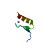


 PDBj
PDBj


 Assembly
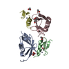


 Mass: 92.094 Da / Num. of mol.: 2 / Source method: obtained synthetically / Formula: C3H8O3
 Mass: 59.044 Da / Num. of mol.: 4 / Source method: obtained synthetically / Formula: C2H3O2
 Mass: 65.409 Da / Num. of mol.: 2 / Source method: obtained synthetically / Formula: Zn
 Sample preparation
 Processing