 Movie
Movie Controller

[English] 日本語
 Yorodumi
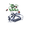
- PDB-1rie: STRUCTURE OF A WATER SOLUBLE FRAGMENT OF THE RIESKE IRON-SULFUR P... -
+ Open data

- Basic information
| Entry | Database: PDB / ID: 1rie | ||||||
|---|---|---|---|---|---|---|---|
| Title | STRUCTURE OF A WATER SOLUBLE FRAGMENT OF THE RIESKE IRON-SULFUR PROTEIN OF THE BOVINE HEART MITOCHONDRIAL CYTOCHROME BC1-COMPLEX | ||||||
 Components Components | RIESKE IRON-SULFUR PROTEIN | ||||||
 Keywords Keywords | ELECTRON TRANSPORT / OXIDOREDUCTASE / CYTOCHROME BC1 COMPLEX / HISTIDINE LIGANDS / RIESKE IRON-SULFUR CLUSTER | ||||||
| Function / homology |  Function and homology information Function and homology informationComplex III assembly / Respiratory electron transport / respiratory chain complex III / quinol-cytochrome-c reductase / quinol-cytochrome-c reductase activity / mitochondrial electron transport, ubiquinol to cytochrome c / 2 iron, 2 sulfur cluster binding / oxidoreductase activity / mitochondrial inner membrane / mitochondrion / metal ion binding Similarity search - Function | ||||||
| Biological species |  | ||||||
| Method |  X-RAY DIFFRACTION / SYNCHROTRON / MAD / Resolution: 1.5 Å X-RAY DIFFRACTION / SYNCHROTRON / MAD / Resolution: 1.5 Å | ||||||
 Authors Authors | Iwata, S. / Saynovits, M. / Link, T.A. / Michel, H. | ||||||
 Citation Citation | Journal: Structure / Year: 1996 Title: Structure of a water soluble fragment of the 'Rieske' iron-sulfur protein of the bovine heart mitochondrial cytochrome bc1 complex determined by MAD phasing at 1.5 A resolution. Authors: Iwata, S. / Saynovits, M. / Link, T.A. / Michel, H. #1: Journal: Eur.J.Biochem. / Year: 1996Title: Isolation, Characterisation and Crystallisation of a Water-Soluble Fragment of the Rieske Iron-Sulfur Protein of Bovine Heart Mitochondrial Bc1 Complex Authors: Link, T.A. / Saynovits, M. / Assmann, C. / Iwata, S. / Ohnishi, T. / Von Jagow, G. #2: Journal: J.Biol.Chem. / Year: 1993Title: The Mitochondrial Targeting Presequence of the Rieske Iron-Sulfur Protein is Processed in a Single Step After Insertion Into the Cytochrome Bc1 Complex in Mammals and Retained as a Subunit in the Complex Authors: Brandt, U. / Yu, L. / Yu, C.A. / Trumpower, B.L. #3: Journal: Biochem.Biophys.Res.Commun. / Year: 1990Title: Cloning and Sequencing of a Cdna Encoding the Rieske Iron-Sulfur Protein of Bovine Heart Mitochondrial Ubiquinol-Cytochrome C Reductase Authors: Usui, S. / Yu, L. / Yu, C.A. #4: Journal: FEBS Lett. / Year: 1987Title: Isolation and Amino Acid Sequence of the 'Rieske' Iron Sulfur Protein of Beef Heart Ubiquinol:Cytochrome C Reductase Authors: Schagger, H. / Borchart, U. / Machleidt, W. / Link, T.A. / Von Jagow, G. | ||||||
| History |
|
- Structure visualization
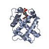
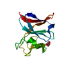
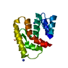
| Structure viewer | Molecule: MolmilJmol/JSmol |
|---|
- Downloads & links
-Download
| PDBx/mmCIF format | 1rie.cif.gz | 41.7 KB | Display | PDBx/mmCIF format |
|---|---|---|---|---|
| PDB format | pdb1rie.ent.gz | 27.7 KB | Display | PDB format |
| PDBx/mmJSON format | 1rie.json.gz | Tree view | PDBx/mmJSON format | |
| Others |  Other downloads Other downloads |
-Validation report
| Arichive directory | https://data.pdbj.org/pub/pdb/validation_reports/ri/1rieftp://data.pdbj.org/pub/pdb/validation_reports/ri/1rie | HTTPS FTP |
|---|
-Related structure data
| Similar structure data |
|---|


-Links
 PDBj
PDBj


- Assembly
| Deposited unit | 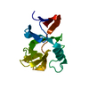
| ||||||||
|---|---|---|---|---|---|---|---|---|---|
| 1 |
| ||||||||
| Unit cell |
|
-Components
| #1: Protein | Mass: 14440.658 Da / Num. of mol.: 1 / Fragment: SOLUBLE FRAGMENT / Source method: isolated from a natural source / Source: (natural) |
|---|---|
| #2: Chemical | ChemComp-FES /   Mass: 175.820 Da / Num. of mol.: 1 / Source method: obtained synthetically / Formula: Fe2S2 Mass: 175.820 Da / Num. of mol.: 1 / Source method: obtained synthetically / Formula: Fe2S2 |
| #3: Water | ChemComp-HOH /  Mass: 18.015 Da / Num. of mol.: 167 / Source method: isolated from a natural source / Formula: H2O Mass: 18.015 Da / Num. of mol.: 167 / Source method: isolated from a natural source / Formula: H2O |
| Has protein modification | Y |
-Experimental details
-Experiment
| Experiment | Method: X-RAY DIFFRACTION / Number of used crystals: 1 |
|---|
- Sample preparation
| Crystal | Density Matthews: 2.2 Å3/Da / Density % sol: 41 % | ||||||||||||||||||||||||||||||
|---|---|---|---|---|---|---|---|---|---|---|---|---|---|---|---|---|---|---|---|---|---|---|---|---|---|---|---|---|---|---|---|
| Crystal grow | pH: 6 / Details: pH 6.0 | ||||||||||||||||||||||||||||||
| Crystal grow | *PLUS Temperature: 22 ℃ / pH: 6.2 / Method: vapor diffusion, hanging drop | ||||||||||||||||||||||||||||||
| Components of the solutions | *PLUS
|
-Data collection
| Diffraction | Mean temperature: 100 K |
|---|---|
| Diffraction source | Source: SYNCHROTRON / Site: Photon Factory  / Beamline: BL-6A / Wavelength: 1 / Beamline: BL-6A / Wavelength: 1 |
| Detector | Type: FUJI / Detector: IMAGE PLATE / Date: May 10, 1995 |
| Radiation | Monochromator: SI(111) / Monochromatic (M) / Laue (L): M / Scattering type: x-ray |
| Radiation wavelength | Wavelength: 1 Å / Relative weight: 1 |
| Reflection | Resolution: 1.5→40 Å / Num. obs: 18058 / % possible obs: 89.4 % / Observed criterion σ(I): -3 / Redundancy: 3.52 % / Rmerge(I) obs: 0.051 / Net I/σ(I): 28.6 |
| Reflection shell | Resolution: 1.5→1.53 Å / Rmerge(I) obs: 0.27 / Mean I/σ(I) obs: 5.5 / % possible all: 68 |
| Reflection | *PLUS Num. measured all: 63633 |
| Reflection shell | *PLUS % possible obs: 68 % |
- Processing
| Software |
| ||||||||||||||||||||||||||||||||||||||||||||||||||||||||||||
|---|---|---|---|---|---|---|---|---|---|---|---|---|---|---|---|---|---|---|---|---|---|---|---|---|---|---|---|---|---|---|---|---|---|---|---|---|---|---|---|---|---|---|---|---|---|---|---|---|---|---|---|---|---|---|---|---|---|---|---|---|---|
| Refinement | Method to determine structure: MAD / Resolution: 1.5→10 Å / σ(F): 2
| ||||||||||||||||||||||||||||||||||||||||||||||||||||||||||||
| Displacement parameters | Biso mean: 9.21 Å2 | ||||||||||||||||||||||||||||||||||||||||||||||||||||||||||||
| Refine analyze | Luzzati sigma a obs: 0.16 Å | ||||||||||||||||||||||||||||||||||||||||||||||||||||||||||||
| Refinement step | Cycle: LAST / Resolution: 1.5→10 Å
| ||||||||||||||||||||||||||||||||||||||||||||||||||||||||||||
| Refine LS restraints |
| ||||||||||||||||||||||||||||||||||||||||||||||||||||||||||||
| Software | *PLUS Name: X-PLOR / Classification: refinement | ||||||||||||||||||||||||||||||||||||||||||||||||||||||||||||
| Refinement | *PLUS Num. reflection all: 15880 / Rfactor all: 0.192 | ||||||||||||||||||||||||||||||||||||||||||||||||||||||||||||
| Solvent computation | *PLUS | ||||||||||||||||||||||||||||||||||||||||||||||||||||||||||||
| Displacement parameters | *PLUS | ||||||||||||||||||||||||||||||||||||||||||||||||||||||||||||
| Refine LS restraints | *PLUS
|
