 Movie
Movie Controller


+ Open data

- Basic information
| Entry | Database: PDB / ID: 1ebt | ||||||
|---|---|---|---|---|---|---|---|
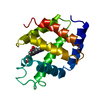
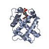
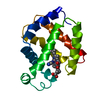
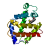
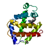
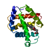
| Title | HEMOGLOBIN I FROM THE CLAM LUCINA PECTINATA BOUND WITH CYANIDE | ||||||
 Components Components | HEMOGLOBIN | ||||||
 Keywords Keywords | OXYGEN TRANSPORT / HEMOGLOBIN / OXYGEN CARRIER / GLOBIN | ||||||
| Function / homology |  Function and homology information Function and homology informationhemoglobin complex / oxygen transport / oxygen binding / heme binding / extracellular region / metal ion binding Similarity search - Function | ||||||
| Biological species |  Lucina pectinata (invertebrata) Lucina pectinata (invertebrata) | ||||||
| Method |  X-RAY DIFFRACTION / MOLECULAR REPLACEMENT / Resolution: 1.9 Å X-RAY DIFFRACTION / MOLECULAR REPLACEMENT / Resolution: 1.9 Å | ||||||
 Authors Authors | Rosano, C. / Bolognesi, M. / Ascenzi, P. | ||||||
 Citation Citation | Journal: Biophys.J. / Year: 1999 Title: Cyanide binding to Lucina pectinata hemoglobin I and to sperm whale myoglobin: an x-ray crystallographic study. Authors: Bolognesi, M. / Rosano, C. / Losso, R. / Borassi, A. / Rizzi, M. / Wittenberg, J.B. / Boffi, A. / Ascenzi, P. | ||||||
| History |
|
- Structure visualization
| Structure viewer | Molecule: MolmilJmol/JSmol |
|---|
- Downloads & links
-Download
| PDBx/mmCIF format | 1ebt.cif.gz | 41.8 KB | Display | PDBx/mmCIF format |
|---|---|---|---|---|
| PDB format | pdb1ebt.ent.gz | 28 KB | Display | PDB format |
| PDBx/mmJSON format | 1ebt.json.gz | Tree view | PDBx/mmJSON format | |
| Others |  Other downloads Other downloads |
-Validation report
| Arichive directory | https://data.pdbj.org/pub/pdb/validation_reports/eb/1ebtftp://data.pdbj.org/pub/pdb/validation_reports/eb/1ebt | HTTPS FTP |
|---|
-Related structure data
| Related structure data | 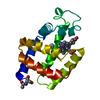 1b0bC  1ebcC  1flpS S: Starting model for refinement C: citing same article ( |
|---|---|
| Similar structure data |


-Links
 PDBj
PDBj


- Assembly
| Deposited unit | 
| ||||||||
|---|---|---|---|---|---|---|---|---|---|
| 1 |
| ||||||||
| Unit cell |
|
-Components
| #1: Protein | Mass: 14744.429 Da / Num. of mol.: 1 / Source method: isolated from a natural source / Source: (natural) Lucina pectinata (invertebrata) / References: UniProt: P41260 |
|---|---|
| #2: Chemical | ChemComp-CYN /   Mass: 26.017 Da / Num. of mol.: 1 / Source method: obtained synthetically / Formula: CN Mass: 26.017 Da / Num. of mol.: 1 / Source method: obtained synthetically / Formula: CN |
| #3: Chemical | ChemComp-HEM /   Mass: 616.487 Da / Num. of mol.: 1 / Source method: obtained synthetically / Formula: C34H32FeN4O4 Mass: 616.487 Da / Num. of mol.: 1 / Source method: obtained synthetically / Formula: C34H32FeN4O4 |
| #4: Water | ChemComp-HOH /  Mass: 18.015 Da / Num. of mol.: 79 / Source method: isolated from a natural source / Formula: H2O Mass: 18.015 Da / Num. of mol.: 79 / Source method: isolated from a natural source / Formula: H2O |
-Experimental details
-Experiment
| Experiment | Method: X-RAY DIFFRACTION / Number of used crystals: 1 |
|---|
- Sample preparation
| Crystal | Density Matthews: 2.5 Å3/Da / Density % sol: 50.8 % | |||||||||||||||
|---|---|---|---|---|---|---|---|---|---|---|---|---|---|---|---|---|
| Crystal grow | pH: 7 / Details: pH 7.00 | |||||||||||||||
| Crystal grow | *PLUS pH: 5.75 / Method: unknown | |||||||||||||||
| Components of the solutions | *PLUS
|
-Data collection
| Diffraction | Mean temperature: 300 K |
|---|---|
| Diffraction source | Source: ROTATING ANODE / Type: RIGAKU RUH2R / Wavelength: 1.5418 |
| Detector | Type: RIGAKU RAXIS / Detector: IMAGE PLATE / Date: Dec 1, 1995 |
| Radiation | Protocol: SINGLE WAVELENGTH / Monochromatic (M) / Laue (L): M / Scattering type: x-ray |
| Radiation wavelength | Wavelength: 1.5418 Å / Relative weight: 1 |
| Reflection | Resolution: 1.9→24 Å / Num. obs: 12085 / % possible obs: 93 % / Redundancy: 2.2 % |
| Reflection | *PLUS Highest resolution: 1.9 Å / Lowest resolution: 24 Å / % possible obs: 93.3 % / Redundancy: 4.1 % / Rmerge(I) obs: 0.054 |
| Reflection shell | *PLUS Highest resolution: 1.9 Å / Lowest resolution: 2 Å / Mean I/σ(I) obs: 5.1 |
- Processing
| Software |
| ||||||||||||||||||||||||||||||||||||||||||||||||||
|---|---|---|---|---|---|---|---|---|---|---|---|---|---|---|---|---|---|---|---|---|---|---|---|---|---|---|---|---|---|---|---|---|---|---|---|---|---|---|---|---|---|---|---|---|---|---|---|---|---|---|---|
| Refinement | Method to determine structure: MOLECULAR REPLACEMENT Starting model: 1FLP Resolution: 1.9→24 Å / Isotropic thermal model: TNT BCORREL / σ(F): 0 / Stereochemistry target values: TNT PROTGEO /
| ||||||||||||||||||||||||||||||||||||||||||||||||||
| Solvent computation | Bsol: 57.8 Å2 / ksol: 0.84 e/Å3 | ||||||||||||||||||||||||||||||||||||||||||||||||||
| Refinement step | Cycle: LAST / Resolution: 1.9→24 Å
| ||||||||||||||||||||||||||||||||||||||||||||||||||
| Refine LS restraints |
| ||||||||||||||||||||||||||||||||||||||||||||||||||
| Software | *PLUS Name: TNT / Version: 5E / Classification: refinement | ||||||||||||||||||||||||||||||||||||||||||||||||||
| Refinement | *PLUS Rfactor obs: 0.184 | ||||||||||||||||||||||||||||||||||||||||||||||||||
| Solvent computation | *PLUS | ||||||||||||||||||||||||||||||||||||||||||||||||||
| Displacement parameters | *PLUS | ||||||||||||||||||||||||||||||||||||||||||||||||||
| Refine LS restraints | *PLUS
|
