 Movie
Movie Controller


+ Open data

- Basic information
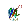
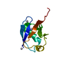
| Entry | Database: PDB / ID: 1ubq | ||||||
|---|---|---|---|---|---|---|---|
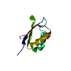
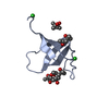
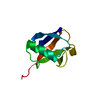
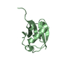
| Title | STRUCTURE OF UBIQUITIN REFINED AT 1.8 ANGSTROMS RESOLUTION | ||||||
 Components Components | UBIQUITIN | ||||||
 Keywords Keywords | CHROMOSOMAL PROTEIN | ||||||
| Function / homology |  Function and homology information Function and homology information: / : / protein modification process => GO:0036211 / : / Peptide chain elongation / Selenocysteine synthesis / Formation of a pool of free 40S subunits / Eukaryotic Translation Termination / SRP-dependent cotranslational protein targeting to membrane / Response of EIF2AK4 (GCN2) to amino acid deficiency ...: / : / protein modification process => GO:0036211 / : / Peptide chain elongation / Selenocysteine synthesis / Formation of a pool of free 40S subunits / Eukaryotic Translation Termination / SRP-dependent cotranslational protein targeting to membrane / Response of EIF2AK4 (GCN2) to amino acid deficiency / Viral mRNA Translation / Nonsense Mediated Decay (NMD) independent of the Exon Junction Complex (EJC) / GTP hydrolysis and joining of the 60S ribosomal subunit / L13a-mediated translational silencing of Ceruloplasmin expression / Major pathway of rRNA processing in the nucleolus and cytosol / Nonsense Mediated Decay (NMD) enhanced by the Exon Junction Complex (EJC) / Maturation of protein E / Maturation of protein E / ER Quality Control Compartment (ERQC) / Myoclonic epilepsy of Lafora / FLT3 signaling by CBL mutants / IRAK2 mediated activation of TAK1 complex / Alpha-protein kinase 1 signaling pathway / Glycogen synthesis / IRAK1 recruits IKK complex / IRAK1 recruits IKK complex upon TLR7/8 or 9 stimulation / Prevention of phagosomal-lysosomal fusion / Endosomal Sorting Complex Required For Transport (ESCRT) / Membrane binding and targetting of GAG proteins / Regulation of TBK1, IKKε (IKBKE)-mediated activation of IRF3, IRF7 / Negative regulation of FLT3 / PTK6 Regulates RTKs and Their Effectors AKT1 and DOK1 / Regulation of TBK1, IKKε-mediated activation of IRF3, IRF7 upon TLR3 ligation / IRAK2 mediated activation of TAK1 complex upon TLR7/8 or 9 stimulation / Constitutive Signaling by NOTCH1 HD Domain Mutants / NOTCH2 Activation and Transmission of Signal to the Nucleus / TICAM1,TRAF6-dependent induction of TAK1 complex / TICAM1-dependent activation of IRF3/IRF7 / APC/C:Cdc20 mediated degradation of Cyclin B / Downregulation of ERBB4 signaling / APC-Cdc20 mediated degradation of Nek2A / Regulation of FZD by ubiquitination / p75NTR recruits signalling complexes / InlA-mediated entry of Listeria monocytogenes into host cells / TRAF6 mediated IRF7 activation in TLR7/8 or 9 signaling / NF-kB is activated and signals survival / TRAF6-mediated induction of TAK1 complex within TLR4 complex / Regulation of pyruvate metabolism / Pexophagy / Downregulation of ERBB2:ERBB3 signaling / Regulation of innate immune responses to cytosolic DNA / NRIF signals cell death from the nucleus / Regulation of PTEN localization / VLDLR internalisation and degradation / cytosolic ribosome / Activated NOTCH1 Transmits Signal to the Nucleus / Synthesis of active ubiquitin: roles of E1 and E2 enzymes / Translesion synthesis by REV1 / TICAM1, RIP1-mediated IKK complex recruitment / Regulation of BACH1 activity / Translesion synthesis by POLK / JNK (c-Jun kinases) phosphorylation and activation mediated by activated human TAK1 / InlB-mediated entry of Listeria monocytogenes into host cell / MAP3K8 (TPL2)-dependent MAPK1/3 activation / Activation of IRF3, IRF7 mediated by TBK1, IKKε (IKBKE) / Downregulation of TGF-beta receptor signaling / Translesion synthesis by POLI / Josephin domain DUBs / Gap-filling DNA repair synthesis and ligation in GG-NER / IKK complex recruitment mediated by RIP1 / PINK1-PRKN Mediated Mitophagy / TGF-beta receptor signaling in EMT (epithelial to mesenchymal transition) / TNFR1-induced NF-kappa-B signaling pathway / Regulation of activated PAK-2p34 by proteasome mediated degradation / TCF dependent signaling in response to WNT / Regulation of NF-kappa B signaling / activated TAK1 mediates p38 MAPK activation / Autodegradation of Cdh1 by Cdh1:APC/C / APC/C:Cdc20 mediated degradation of Securin / NOTCH3 Activation and Transmission of Signal to the Nucleus / Regulation of signaling by CBL / Negative regulators of DDX58/IFIH1 signaling / N-glycan trimming in the ER and Calnexin/Calreticulin cycle / Asymmetric localization of PCP proteins / Fanconi Anemia Pathway / Negative regulation of FGFR3 signaling / Ubiquitin-dependent degradation of Cyclin D / Peroxisomal protein import / Deactivation of the beta-catenin transactivating complex / SCF-beta-TrCP mediated degradation of Emi1 / NIK-->noncanonical NF-kB signaling / Stabilization of p53 / AUF1 (hnRNP D0) binds and destabilizes mRNA / TNFR2 non-canonical NF-kB pathway / Negative regulation of FGFR2 signaling / Negative regulation of FGFR4 signaling / Downregulation of SMAD2/3:SMAD4 transcriptional activity / Negative regulation of FGFR1 signaling / Termination of translesion DNA synthesis / Assembly of the pre-replicative complex Similarity search - Function | ||||||
| Biological species |  Homo sapiens (human) Homo sapiens (human) | ||||||
| Method |  X-RAY DIFFRACTION / Resolution: 1.8 Å X-RAY DIFFRACTION / Resolution: 1.8 Å | ||||||
 Authors Authors | Vijay-Kumar, S. / Bugg, C.E. / Cook, W.J. | ||||||
 Citation Citation | Journal: J Mol Biol / Year: 1987 Title: Structure of ubiquitin refined at 1.8 A resolution. Abstract: The crystal structure of human erythrocytic ubiquitin has been refined at 1.8 A resolution using a restrained least-squares procedure. The crystallographic R-factor for the final model is 0.176. Bond ...The crystal structure of human erythrocytic ubiquitin has been refined at 1.8 A resolution using a restrained least-squares procedure. The crystallographic R-factor for the final model is 0.176. Bond lengths and bond angles in the molecule have root-mean-square deviations from ideal values of 0.016 A and 1.5 degrees, respectively. A total of 58 water molecules per molecule of ubiquitin are included in the final model. The last four residues in the molecule appear to have partial occupancy or large thermal motion. The overall structure of ubiquitin is extremely compact and tightly hydrogen-bonded; approximately 87% of the polypeptide chain is involved in hydrogen-bonded secondary structure. Prominent secondary structural features include three and one-half turns of alpha-helix, a short piece of 3(10)-helix, a mixed beta-sheet that contains five strands, and seven reverse turns. There is a marked hydrophobic core formed between the beta-sheet and alpha-helix. The molecule features a number of unusual secondary structural features, including a parallel G1 beta-bulge, two reverse Asx turns, and a symmetrical hydrogen-bonding region that involves the two helices and two of the reverse turns. #1: Journal: J.Biol.Chem. / Year: 1987Title: Comparison of the Three-Dimensional Structures of Human, Yeast, and Oat Ubiquitin Authors: Vijay-Kumar, S. / Bugg, C.E. / Wilkinson, K.D. / Vierstra, R.D. / Hatfield, P.M. / Cook, W.J. #2: Journal: Proc.Natl.Acad.Sci.USA / Year: 1985Title: Three-Dimensional Structure of Ubiquitin at 2.8 Angstroms Resolution Authors: Vijay-Kumar, S. / Bugg, C.E. / Wilkinson, K.D. / Cook, W.J. #3: Journal: J.Mol.Biol. / Year: 1979Title: Crystallization and Preliminary X-Ray Investigation of Ubiquitin, a Non-Histone Chromosomal Protein Authors: Cook, W.J. / Suddath, F.L. / Bugg, C.E. / Goldstein, G. #4: Journal: Nature / Year: 1975Title: Molecular Conservation of 74 Amino Acid Sequence of Ubiquitin between Cattle and Man Authors: Schlesinger, D.H. / Goldstein, G. | ||||||
| History |
|
- Structure visualization
| Structure viewer | Molecule: MolmilJmol/JSmol |
|---|
- Downloads & links
-Download
| PDBx/mmCIF format | 1ubq.cif.gz | 26.3 KB | Display | PDBx/mmCIF format |
|---|---|---|---|---|
| PDB format | pdb1ubq.ent.gz | 17.5 KB | Display | PDB format |
| PDBx/mmJSON format | 1ubq.json.gz | Tree view | PDBx/mmJSON format | |
| Others |  Other downloads Other downloads |
-Validation report
| Arichive directory | https://data.pdbj.org/pub/pdb/validation_reports/ub/1ubqftp://data.pdbj.org/pub/pdb/validation_reports/ub/1ubq | HTTPS FTP |
|---|
-Related structure data
| Similar structure data |
|---|


-Links
 PDBj
PDBj


- Assembly
| Deposited unit | 
| ||||||||
|---|---|---|---|---|---|---|---|---|---|
| 1 |
| ||||||||
| Unit cell |
|
-Components
| #1: Protein | Mass: 8576.831 Da / Num. of mol.: 1 Source method: isolated from a genetically manipulated source Source: (gene. exp.) Homo sapiens (human) / References: UniProt: P62988, UniProt: P0CG48*PLUS |
|---|---|
| #2: Water | ChemComp-HOH /  Mass: 18.015 Da / Num. of mol.: 58 / Source method: isolated from a natural source / Formula: H2O Mass: 18.015 Da / Num. of mol.: 58 / Source method: isolated from a natural source / Formula: H2O |
-Experimental details
-Experiment
| Experiment | Method: X-RAY DIFFRACTION |
|---|
- Sample preparation
| Crystal | Density Matthews: 1.83 Å3/Da / Density % sol: 32.94 % |
|---|---|
| Crystal grow | *PLUS Method: other / Details: Cook, W.J., (1979) J. Mol. Biol., 130, 353. |
-Data collection
| Radiation | Scattering type: x-ray |
|---|---|
| Radiation wavelength | Relative weight: 1 |
| Reflection | *PLUS Highest resolution: 1.8 Å / Lowest resolution: 6 Å / Num. all: 5750 / Num. obs: 5554 / Observed criterion σ(I): 2.5 / Num. measured all: 6029 |
- Processing
| Software | Name: PROLSQ / Classification: refinement | ||||||||||||
|---|---|---|---|---|---|---|---|---|---|---|---|---|---|
| Refinement | Rfactor obs: 0.176 / Highest resolution: 1.8 Å | ||||||||||||
| Refinement step | Cycle: LAST / Highest resolution: 1.8 Å
| ||||||||||||
| Refine LS restraints |
| ||||||||||||
| Refinement | *PLUS Lowest resolution: 6 Å / Num. reflection obs: 5554 / σ(I): 2.5 | ||||||||||||
| Solvent computation | *PLUS | ||||||||||||
| Displacement parameters | *PLUS |
