 Movie
Movie Controller


+ Open data

- Basic information
| Entry | Database: PDB / ID: 1jnl | ||||||
|---|---|---|---|---|---|---|---|
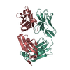
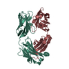
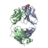
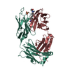
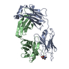
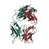
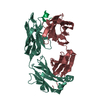
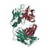
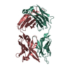
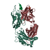
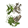
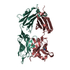
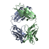
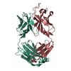
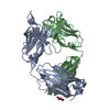
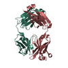
| Title | Crystal Structure of Fab-Estradiol Complexes | ||||||
 Components Components |
| ||||||
 Keywords Keywords | IMMUNE SYSTEM / IGG FOLD / ANTIBODY-HAPTEN COMPLEX / ESTRADIOL | ||||||
| Function / homology |  Function and homology information Function and homology informationimmunoglobulin complex / adaptive immune response / extracellular region / metal ion binding / plasma membrane Similarity search - Function | ||||||
| Biological species |  | ||||||
| Method |  X-RAY DIFFRACTION / SYNCHROTRON / MOLECULAR REPLACEMENT / Resolution: 3 Å X-RAY DIFFRACTION / SYNCHROTRON / MOLECULAR REPLACEMENT / Resolution: 3 Å | ||||||
 Authors Authors | Monnet, C. / Bettsworth, F. / Stura, E.A. / Le Du, M.-H. / Menez, R. / Derrien, L. / Zinn-Justin, S. / Gilquin, B. / Sibai, G. / Battail-Poirot, N. ...Monnet, C. / Bettsworth, F. / Stura, E.A. / Le Du, M.-H. / Menez, R. / Derrien, L. / Zinn-Justin, S. / Gilquin, B. / Sibai, G. / Battail-Poirot, N. / Jolivet, M. / Menez, A. / Arnaud, M. / Ducancel, F. / Charbonnier, J.B. | ||||||
 Citation Citation | Journal: J.Mol.Biol. / Year: 2002 Title: Highly specific anti-estradiol antibodies: structural characterisation and binding diversity. Authors: Monnet, C. / Bettsworth, F. / Stura, E.A. / Du, M.H. / Menez, R. / Derrien, L. / Zinn-Justin, S. / Gilquin, B. / Sibai, G. / Battail-Poirot, N. / Jolivet, M. / Menez, A. / Arnaud, M. / ...Authors: Monnet, C. / Bettsworth, F. / Stura, E.A. / Du, M.H. / Menez, R. / Derrien, L. / Zinn-Justin, S. / Gilquin, B. / Sibai, G. / Battail-Poirot, N. / Jolivet, M. / Menez, A. / Arnaud, M. / Ducancel, F. / Charbonnier, J.B. | ||||||
| History |
|
- Structure visualization
| Structure viewer | Molecule: MolmilJmol/JSmol |
|---|
- Downloads & links
-Download
| PDBx/mmCIF format | 1jnl.cif.gz | 83.6 KB | Display | PDBx/mmCIF format |
|---|---|---|---|---|
| PDB format | pdb1jnl.ent.gz | 63.6 KB | Display | PDB format |
| PDBx/mmJSON format | 1jnl.json.gz | Tree view | PDBx/mmJSON format | |
| Others |  Other downloads Other downloads |
-Validation report
| Arichive directory | https://data.pdbj.org/pub/pdb/validation_reports/jn/1jnlftp://data.pdbj.org/pub/pdb/validation_reports/jn/1jnl | HTTPS FTP |
|---|
-Related structure data
| Related structure data | 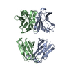 1jn6C  1jnhC  1jnnC  1bbdS S: Starting model for refinement C: citing same article ( |
|---|---|
| Similar structure data |


-Links
 PDBj
PDBj


- Assembly
| Deposited unit | 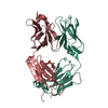
| ||||||||
|---|---|---|---|---|---|---|---|---|---|
| 1 |
| ||||||||
| Unit cell |
|
-Components
| #1: Antibody | Mass: 23426.963 Da / Num. of mol.: 1 / Source method: isolated from a natural source / Source: (natural) |
|---|---|
| #2: Antibody | Mass: 23162.977 Da / Num. of mol.: 1 / Source method: isolated from a natural source / Source: (natural) |
| Has protein modification | Y |
-Experimental details
-Experiment
| Experiment | Method: X-RAY DIFFRACTION / Number of used crystals: 1 |
|---|
- Sample preparation
| Crystal | Density Matthews: 2.91 Å3/Da / Density % sol: 57.71 % | ||||||||||||||||||||||||||||||
|---|---|---|---|---|---|---|---|---|---|---|---|---|---|---|---|---|---|---|---|---|---|---|---|---|---|---|---|---|---|---|---|
| Crystal grow | Temperature: 298 K / Method: vapor diffusion, sitting drop / pH: 9.3 Details: PEG 8000, CHES, pH 9.3, VAPOR DIFFUSION, SITTING DROP, temperature 298K | ||||||||||||||||||||||||||||||
| Crystal grow | *PLUS pH: 7 / Method: vapor diffusion | ||||||||||||||||||||||||||||||
| Components of the solutions | *PLUS
|
-Data collection
| Diffraction | Mean temperature: 279 K |
|---|---|
| Diffraction source | Source: SYNCHROTRON / Site: LURE  / Beamline: DW32 / Wavelength: 1 Å / Beamline: DW32 / Wavelength: 1 Å |
| Detector | Type: MARRESEARCH / Detector: IMAGE PLATE / Date: Jul 17, 1997 |
| Radiation | Protocol: SINGLE WAVELENGTH / Monochromatic (M) / Laue (L): M / Scattering type: x-ray |
| Radiation wavelength | Wavelength: 1 Å / Relative weight: 1 |
| Reflection | Resolution: 3→20 Å / Num. all: 10882 / Num. obs: 10863 / % possible obs: 96.8 % / Observed criterion σ(F): 2 / Observed criterion σ(I): 2 / Redundancy: 6.5 % / Biso Wilson estimate: 45.2 Å2 / Rsym value: 0.087 / Net I/σ(I): 9.2 |
| Reflection shell | Resolution: 3→3.1 Å / Mean I/σ(I) obs: 2.4 / Rsym value: 0.291 / % possible all: 96.2 |
- Processing
| Software |
| ||||||||||||||||||||
|---|---|---|---|---|---|---|---|---|---|---|---|---|---|---|---|---|---|---|---|---|---|
| Refinement | Method to determine structure: MOLECULAR REPLACEMENT Starting model: PDB entry: 1bbd Resolution: 3→20 Å / σ(F): 2 / Stereochemistry target values: Engh & Huber
| ||||||||||||||||||||
| Refine analyze | Luzzati sigma a obs: 0.42 Å | ||||||||||||||||||||
| Refinement step | Cycle: LAST / Resolution: 3→20 Å
| ||||||||||||||||||||
| Refine LS restraints |
| ||||||||||||||||||||
| Software | *PLUS Name: CNS / Classification: refinement | ||||||||||||||||||||
| Refinement | *PLUS Highest resolution: 3 Å / Lowest resolution: 10 Å / σ(F): 2 / Rfactor Rfree: 0.29 | ||||||||||||||||||||
| Solvent computation | *PLUS | ||||||||||||||||||||
| Displacement parameters | *PLUS | ||||||||||||||||||||
| Refine LS restraints | *PLUS
|
