 Movie
Movie Controller

[English] 日本語
 Yorodumi
- PDB-2ajz: Crystal Structure of Cocaine catalytic Antibody 7A1 Fab' in Compl... -
+ Open data

- Basic information
| Entry | Database: PDB / ID: 2ajz | ||||||
|---|---|---|---|---|---|---|---|
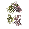
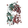
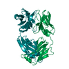
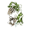
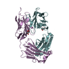
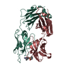
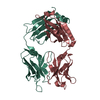
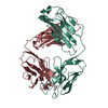
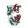
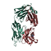
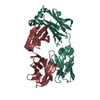
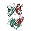
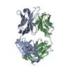
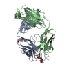
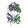
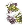
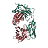
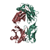
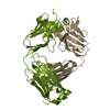
| Title | Crystal Structure of Cocaine catalytic Antibody 7A1 Fab' in Complex with ecgonine methyl ester | ||||||
 Components Components | (Antibody 7A1 Fab') x 2 | ||||||
 Keywords Keywords | IMMUNE SYSTEM / catalytic Antibody / Fab / ecgonine methyl ester / HYDROLYTIC | ||||||
| Function / homology |  Function and homology information Function and homology informationalpha-beta T cell receptor complex / IgG immunoglobulin complex / immunoglobulin complex / B cell differentiation / adaptive immune response / extracellular region / plasma membrane Similarity search - Function | ||||||
| Biological species |  | ||||||
| Method |  X-RAY DIFFRACTION / SYNCHROTRON / MOLECULAR REPLACEMENT / Resolution: 2.3 Å X-RAY DIFFRACTION / SYNCHROTRON / MOLECULAR REPLACEMENT / Resolution: 2.3 Å | ||||||
 Authors Authors | Zhu, X. / Wilson, I.A. | ||||||
 Citation Citation | Journal: Structure / Year: 2006 Title: Complete reaction cycle of a cocaine catalytic antibody at atomic resolution. Authors: Zhu, X. / Dickerson, T.J. / Rogers, C.J. / Kaufmann, G.F. / Mee, J.M. / McKenzie, K.M. / Janda, K.D. / Wilson, I.A. | ||||||
| History |
| ||||||
| Remark 999 | SEQUENCE The sequence of these proteins are not available at any sequence database at the time of processing. |
- Structure visualization
| Structure viewer | Molecule: MolmilJmol/JSmol |
|---|
- Downloads & links
-Download
| PDBx/mmCIF format | 2ajz.cif.gz | 183 KB | Display | PDBx/mmCIF format |
|---|---|---|---|---|
| PDB format | pdb2ajz.ent.gz | 145 KB | Display | PDB format |
| PDBx/mmJSON format | 2ajz.json.gz | Tree view | PDBx/mmJSON format | |
| Others |  Other downloads Other downloads |
-Validation report
| Arichive directory | https://data.pdbj.org/pub/pdb/validation_reports/aj/2ajzftp://data.pdbj.org/pub/pdb/validation_reports/aj/2ajz | HTTPS FTP |
|---|
-Related structure data
| Related structure data | 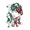 2ajsSC 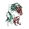 2ajuC 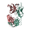 2ajvC  2ajxC 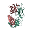 2ajyC 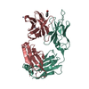 2ak1C S: Starting model for refinement C: citing same article ( |
|---|---|
| Similar structure data |

-Links
 PDBj
PDBj


- Assembly
| Deposited unit | 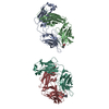
| ||||||||
|---|---|---|---|---|---|---|---|---|---|
| 1 | 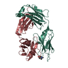
| ||||||||
| 2 | 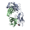
| ||||||||
| Unit cell |
|
-Components
| #1: Antibody | Mass: 24059.611 Da / Num. of mol.: 2 / Fragment: IMMUNOGLOBULIN IGG1 KAPPA LIGHT CHAIN / Source method: isolated from a natural source / Source: (natural) #2: Antibody | Mass: 23865.783 Da / Num. of mol.: 2 / Fragment: IMMUNOGLOBULIN IGG1 heavy CHAIN / Source method: isolated from a natural source / Source: (natural) #3: Chemical |   Mass: 199.247 Da / Num. of mol.: 2 / Source method: obtained synthetically / Formula: C10H17NO3 Mass: 199.247 Da / Num. of mol.: 2 / Source method: obtained synthetically / Formula: C10H17NO3#4: Water | ChemComp-HOH / |  Mass: 18.015 Da / Num. of mol.: 263 / Source method: isolated from a natural source / Formula: H2O Mass: 18.015 Da / Num. of mol.: 263 / Source method: isolated from a natural source / Formula: H2OHas protein modification | Y | |
|---|
-Experimental details
-Experiment
| Experiment | Method: X-RAY DIFFRACTION / Number of used crystals: 1 |
|---|
- Sample preparation
| Crystal | Density Matthews: 2.6 Å3/Da / Density % sol: 52.7 % |
|---|---|
| Crystal grow | Temperature: 295 K / Method: vapor diffusion, sitting drop / pH: 5.5 Details: PEG 4000, ammonium fluoride, sodium acetate, pH 5.5, VAPOR DIFFUSION, SITTING DROP, temperature 295K |
-Data collection
| Diffraction | Mean temperature: 100 K |
|---|---|
| Diffraction source | Source: SYNCHROTRON / Site: ALS  / Beamline: 5.0.2 / Wavelength: 1 Å / Beamline: 5.0.2 / Wavelength: 1 Å |
| Detector | Type: ADSC QUANTUM 210 / Detector: CCD / Date: Aug 8, 2003 |
| Radiation | Monochromator: DOUBLE-CRYSTAL, Si(111) / Protocol: SINGLE WAVELENGTH / Monochromatic (M) / Laue (L): M / Scattering type: x-ray |
| Radiation wavelength | Wavelength: 1 Å / Relative weight: 1 |
| Reflection | Resolution: 2.3→50 Å / Num. obs: 40412 / % possible obs: 90.2 % / Rmerge(I) obs: 0.099 / Χ2: 3.303 |
| Reflection shell | Resolution: 2.3→2.38 Å / % possible obs: 65.2 % / Rmerge(I) obs: 0.363 / Num. measured obs: 2905 / Χ2: 3.576 / % possible all: 65.2 |
- Processing
| Software |
| ||||||||||||||||||||||||||||||||
|---|---|---|---|---|---|---|---|---|---|---|---|---|---|---|---|---|---|---|---|---|---|---|---|---|---|---|---|---|---|---|---|---|---|
| Refinement | Method to determine structure: MOLECULAR REPLACEMENT Starting model: PDb ENTRY 2AJS Resolution: 2.3→50 Å / Cross valid method: THROUGHOUT / σ(F): 0 / Stereochemistry target values: Engh & Huber
| ||||||||||||||||||||||||||||||||
| Solvent computation | Bsol: 60.308 Å2 | ||||||||||||||||||||||||||||||||
| Displacement parameters | Biso mean: 35.657 Å2
| ||||||||||||||||||||||||||||||||
| Refinement step | Cycle: LAST / Resolution: 2.3→50 Å
| ||||||||||||||||||||||||||||||||
| Refine LS restraints |
|
