 Movie
Movie Controller


+ Open data

- Basic information
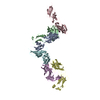
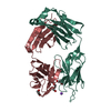
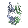
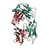
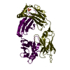
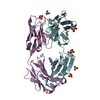
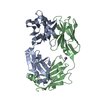
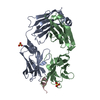
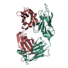
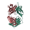
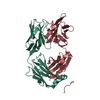
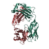
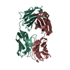
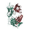
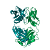
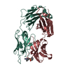
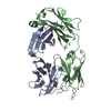
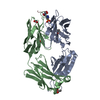
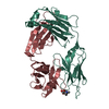
| Entry | Database: PDB / ID: 1fgn | ||||||
|---|---|---|---|---|---|---|---|
| Title | MONOCLONAL MURINE ANTIBODY 5G9-ANTI-HUMAN TISSUE FACTOR | ||||||
 Components Components | (IMMUNOGLOBULIN FAB 5G9) x 2 | ||||||
 Keywords Keywords | IMMUNOGLOBULIN / ANTIBODY / FAB / ANTI-TF / MONOCLONAL / MURINE | ||||||
| Function / homology |  Function and homology information Function and homology informationalpha-beta T cell receptor complex / IgG immunoglobulin complex / B cell differentiation / adaptive immune response / extracellular region / plasma membrane Similarity search - Function | ||||||
| Biological species |  | ||||||
| Method |  X-RAY DIFFRACTION / MOLECULAR REPLACEMENT / Resolution: 2.5 Å X-RAY DIFFRACTION / MOLECULAR REPLACEMENT / Resolution: 2.5 Å | ||||||
 Authors Authors | Huang, M. / Syed, R. / Stura, E.A. / Stone, M.J. / Stefanko, R.S. / Ruf, W. / Edgington, T.S. / Wilson, I.A. | ||||||
 Citation Citation | Journal: J.Mol.Biol. / Year: 1998 Title: The mechanism of an inhibitory antibody on TF-initiated blood coagulation revealed by the crystal structures of human tissue factor, Fab 5G9 and TF.5G9 complex. Authors: Huang, M. / Syed, R. / Stura, E.A. / Stone, M.J. / Stefanko, R.S. / Ruf, W. / Edgington, T.S. / Wilson, I.A. | ||||||
| History |
|
- Structure visualization
| Structure viewer | Molecule: MolmilJmol/JSmol |
|---|
- Downloads & links
-Download
| PDBx/mmCIF format | 1fgn.cif.gz | 92.4 KB | Display | PDBx/mmCIF format |
|---|---|---|---|---|
| PDB format | pdb1fgn.ent.gz | 69.7 KB | Display | PDB format |
| PDBx/mmJSON format | 1fgn.json.gz | Tree view | PDBx/mmJSON format | |
| Others |  Other downloads Other downloads |
-Validation report
| Arichive directory | https://data.pdbj.org/pub/pdb/validation_reports/fg/1fgnftp://data.pdbj.org/pub/pdb/validation_reports/fg/1fgn | HTTPS FTP |
|---|
-Related structure data


-Links
 PDBj
PDBj


- Assembly
| Deposited unit | 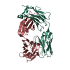
| ||||||||
|---|---|---|---|---|---|---|---|---|---|
| 1 |
| ||||||||
| Unit cell |
|
-Components
| #1: Antibody | Mass: 23810.203 Da / Num. of mol.: 1 Fragment: LIGHT CHAIN RESIDUES 1 - 214, HEAVY CHAIN RESIDUES 1 - 214 Source method: isolated from a natural source / Details: A MONOCLONAL FAB AGAINST HUMAN TISSUE FACTOR / Source: (natural) |
|---|---|
| #2: Antibody | Mass: 23175.799 Da / Num. of mol.: 1 Fragment: LIGHT CHAIN RESIDUES 1 - 214, HEAVY CHAIN RESIDUES 1 - 214 Source method: isolated from a natural source / Details: A MONOCLONAL FAB AGAINST HUMAN TISSUE FACTOR / Source: (natural) |
| Has protein modification | Y |
-Experimental details
-Experiment
| Experiment | Method: X-RAY DIFFRACTION / Number of used crystals: 3 |
|---|
- Sample preparation
| Crystal | Density Matthews: 2.4 Å3/Da / Density % sol: 46 % | |||||||||||||||
|---|---|---|---|---|---|---|---|---|---|---|---|---|---|---|---|---|
| Crystal grow | pH: 7 Details: TWO SLIGHTLY DIFFERENT CRYSTAL FORMS OF FAB 5G9 WERE OBTAINED USING 17.5% PEG 10K AT PH 8.5 AND 1.35M SODIUM CITRATE AT PH 5.5 AS PRECIPITANTS. THE TWO CRYSTAL FORMS ARE BOTH ORTHORHOMBIC, ...Details: TWO SLIGHTLY DIFFERENT CRYSTAL FORMS OF FAB 5G9 WERE OBTAINED USING 17.5% PEG 10K AT PH 8.5 AND 1.35M SODIUM CITRATE AT PH 5.5 AS PRECIPITANTS. THE TWO CRYSTAL FORMS ARE BOTH ORTHORHOMBIC, P212121, WITH TYPICAL SIZES OF 0.5 X 0.4 X 0.3 MM3 (CITRATE FORM) AND 0.6 X 0.35 X 0.2 MM3 (PEG FORM), WITH SLIGHTLY DIFFERENT CELL PARAMETERS OF A=89.1A, B=90.5A, C=59.3A (CITRATE) AND A=91.6A, B=93.7A, C=60.7A (PEG). PEG FORM IS USED FOR STRUCTURE DETERMINATION., pH 7.0 | |||||||||||||||
| Crystal grow | *PLUS pH: 8.5 / Method: unknown | |||||||||||||||
| Components of the solutions | *PLUS
|
-Data collection
| Diffraction | Mean temperature: 297 K |
|---|---|
| Diffraction source | Source: ROTATING ANODE / Type: ELLIOTT GX-18 / Wavelength: 1.5418 |
| Detector | Type: SIEMENS / Detector: AREA DETECTOR / Date: Mar 1, 1992 / Details: COLLIMATOR |
| Radiation | Monochromator: NI FILTER / Monochromatic (M) / Laue (L): M / Scattering type: x-ray |
| Radiation wavelength | Wavelength: 1.5418 Å / Relative weight: 1 |
| Reflection | Resolution: 2.38→37 Å / Num. obs: 19394 / % possible obs: 90 % / Observed criterion σ(I): 0 / Redundancy: 4.5 % / Biso Wilson estimate: 30 Å2 / Rsym value: 0.075 / Net I/σ(I): 20 |
| Reflection shell | Resolution: 2.39→2.54 Å / Redundancy: 1.9 % / Mean I/σ(I) obs: 4 / Rsym value: 0.183 / % possible all: 58.5 |
| Reflection | *PLUS Num. measured all: 86379 / Rmerge(I) obs: 0.075 |
| Reflection shell | *PLUS % possible obs: 58.5 % / Rmerge(I) obs: 0.182 / Mean I/σ(I) obs: 2.4 |
- Processing
| Software |
| ||||||||||||||||||||||||||||||||||||||||||||||||||||||||||||||||||||||||||||||||
|---|---|---|---|---|---|---|---|---|---|---|---|---|---|---|---|---|---|---|---|---|---|---|---|---|---|---|---|---|---|---|---|---|---|---|---|---|---|---|---|---|---|---|---|---|---|---|---|---|---|---|---|---|---|---|---|---|---|---|---|---|---|---|---|---|---|---|---|---|---|---|---|---|---|---|---|---|---|---|---|---|---|
| Refinement | Method to determine structure: MOLECULAR REPLACEMENT Starting model: FABS: HYHEL5-5 AND MCPC603 Resolution: 2.5→8 Å / Rfactor Rfree error: 0.005 / Data cutoff high absF: 10000000 / Data cutoff low absF: 0.001 / Isotropic thermal model: RESTRAINED / Cross valid method: A POSTERIORI / σ(F): 0
| ||||||||||||||||||||||||||||||||||||||||||||||||||||||||||||||||||||||||||||||||
| Displacement parameters | Biso mean: 10.2 Å2 | ||||||||||||||||||||||||||||||||||||||||||||||||||||||||||||||||||||||||||||||||
| Refine analyze | Luzzati coordinate error obs: 0.27 Å / Luzzati d res low obs: 5 Å / Luzzati sigma a obs: 0.28 Å | ||||||||||||||||||||||||||||||||||||||||||||||||||||||||||||||||||||||||||||||||
| Refinement step | Cycle: LAST / Resolution: 2.5→8 Å
| ||||||||||||||||||||||||||||||||||||||||||||||||||||||||||||||||||||||||||||||||
| Refine LS restraints |
| ||||||||||||||||||||||||||||||||||||||||||||||||||||||||||||||||||||||||||||||||
| LS refinement shell | Resolution: 2.5→2.61 Å / Rfactor Rfree error: 0.02 / Total num. of bins used: 8
| ||||||||||||||||||||||||||||||||||||||||||||||||||||||||||||||||||||||||||||||||
| Xplor file |
| ||||||||||||||||||||||||||||||||||||||||||||||||||||||||||||||||||||||||||||||||
| Software | *PLUS Name: X-PLOR / Version: 3.1 / Classification: refinement | ||||||||||||||||||||||||||||||||||||||||||||||||||||||||||||||||||||||||||||||||
| Refine LS restraints | *PLUS
|
