 Movie
Movie Controller

[English] 日本語
 Yorodumi
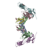
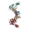
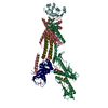
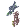
- PDB-1ahw: A COMPLEX OF EXTRACELLULAR DOMAIN OF TISSUE FACTOR WITH AN INHIBI... -
+ Open data

- Basic information
| Entry | Database: PDB / ID: 1ahw | ||||||
|---|---|---|---|---|---|---|---|
| Title | A COMPLEX OF EXTRACELLULAR DOMAIN OF TISSUE FACTOR WITH AN INHIBITORY FAB (5G9) | ||||||
 Components Components |
| ||||||
 Keywords Keywords | COMPLEX (IMMUNOGLOBULIN/TISSUE FACTOR) / BLOOD COAGULATION / TISSUE FACTOR / FAB / COMPLEX / ANTIBODY / COMPLEX (IMMUNOGLOBULIN-TISSUE FACTOR) / COMPLEX (IMMUNOGLOBULIN-TISSUE FACTOR) complex | ||||||
| Function / homology |  Function and homology information Function and homology informationactivation of plasma proteins involved in acute inflammatory response / activation of blood coagulation via clotting cascade / serine-type peptidase complex / positive regulation of platelet-derived growth factor receptor signaling pathway / NGF-stimulated transcription / cytokine receptor activity / positive regulation of positive chemotaxis / alpha-beta T cell receptor complex / : / IgG immunoglobulin complex ...activation of plasma proteins involved in acute inflammatory response / activation of blood coagulation via clotting cascade / serine-type peptidase complex / positive regulation of platelet-derived growth factor receptor signaling pathway / NGF-stimulated transcription / cytokine receptor activity / positive regulation of positive chemotaxis / alpha-beta T cell receptor complex / : / IgG immunoglobulin complex / positive regulation of endothelial cell apoptotic process / immunoglobulin complex / positive regulation of TOR signaling / positive regulation of endothelial cell proliferation / B cell differentiation / positive regulation of interleukin-8 production / protein processing / phospholipid binding / cytokine-mediated signaling pathway / positive regulation of angiogenesis / blood coagulation / extracellular matrix / protease binding / adaptive immune response / positive regulation of cell migration / external side of plasma membrane / positive regulation of gene expression / cell surface / : / extracellular region / membrane / plasma membrane Similarity search - Function | ||||||
| Biological species |  Homo sapiens (human) Homo sapiens (human) | ||||||
| Method |  X-RAY DIFFRACTION / MOLECULAR REPLACEMENT / Resolution: 3 Å X-RAY DIFFRACTION / MOLECULAR REPLACEMENT / Resolution: 3 Å | ||||||
 Authors Authors | Huang, M. / Syed, R. / Stura, E.A. / Stone, M.J. / Stefanko, R.S. / Ruf, W. / Edgington, T.S. / Wilson, I.A. | ||||||
 Citation Citation | Journal: J.Mol.Biol. / Year: 1998 Title: The mechanism of an inhibitory antibody on TF-initiated blood coagulation revealed by the crystal structures of human tissue factor, Fab 5G9 and TF.5G9 complex. Authors: Huang, M. / Syed, R. / Stura, E.A. / Stone, M.J. / Stefanko, R.S. / Ruf, W. / Edgington, T.S. / Wilson, I.A. | ||||||
| History |
|
- Structure visualization
| Structure viewer | Molecule: MolmilJmol/JSmol |
|---|
- Downloads & links
-Download
| PDBx/mmCIF format | 1ahw.cif.gz | 229.1 KB | Display | PDBx/mmCIF format |
|---|---|---|---|---|
| PDB format | pdb1ahw.ent.gz | 186.4 KB | Display | PDB format |
| PDBx/mmJSON format | 1ahw.json.gz | Tree view | PDBx/mmJSON format | |
| Others |  Other downloads Other downloads |
-Validation report
| Arichive directory | https://data.pdbj.org/pub/pdb/validation_reports/ah/1ahwftp://data.pdbj.org/pub/pdb/validation_reports/ah/1ahw | HTTPS FTP |
|---|
-Related structure data
| Related structure data |  1fgnSC  1tfhSC S: Starting model for refinement C: citing same article ( |
|---|---|
| Similar structure data |


-Links
 PDBj
PDBj


- Assembly
| Deposited unit | 
| |||||||||||||||
|---|---|---|---|---|---|---|---|---|---|---|---|---|---|---|---|---|
| 1 |
| |||||||||||||||
| Unit cell |
| |||||||||||||||
| Noncrystallographic symmetry (NCS) | NCS domain:
NCS oper: (Code: given Matrix: (-0.253131, -0.068772, 0.964984), Vector: |
-Components
| #1: Antibody | Mass: 23810.203 Da / Num. of mol.: 2 / Fragment: LIGHT CHAIN RESIDUES 1 - 214 / Source method: isolated from a natural source / Source: (natural) #2: Antibody | Mass: 23175.799 Da / Num. of mol.: 2 / Fragment: HEAVY CHAIN RESIDUES 1 - 214 / Source method: isolated from a natural source / Source: (natural) #3: Protein | Mass: 24826.512 Da / Num. of mol.: 2 / Fragment: EXTRACELLULAR DOMAIN Source method: isolated from a genetically manipulated source Source: (gene. exp.) Homo sapiens (human) / Cell line: BL21 / Gene: HUMAN TISSUE FACTOR EXTRACELLU / Organ: BLOOD / Plasmid: PTRCHISC (INVITROGEN) / Species (production host): Escherichia coli / Cellular location (production host): INCLUSION BODIESGene (production host): HUMAN TISSUE FACTOR EXTRACELLULAR DOMAIN Production host:  Has protein modification | Y | |
|---|
-Experimental details
-Experiment
| Experiment | Method: X-RAY DIFFRACTION / Number of used crystals: 4 |
|---|
- Sample preparation
| Crystal | Density Matthews: 3.7 Å3/Da / Density % sol: 64 % | |||||||||||||||||||||||||
|---|---|---|---|---|---|---|---|---|---|---|---|---|---|---|---|---|---|---|---|---|---|---|---|---|---|---|
| Crystal grow | pH: 7 Details: TF-5G9 CRYSTAL WERE GROWN IN 1.7-2.0M AMMONIUM SULFATE, 0.1M SODIUM CITRATE, PH 5.0-5.5, 0.2% 2-METHYL-2,4-PENTANE-DIOL (MPD), AND 2% PEG 600 AT AN EQUIMOLAR 5G9:TF RATIO. THE CRYSTALS GREW ...Details: TF-5G9 CRYSTAL WERE GROWN IN 1.7-2.0M AMMONIUM SULFATE, 0.1M SODIUM CITRATE, PH 5.0-5.5, 0.2% 2-METHYL-2,4-PENTANE-DIOL (MPD), AND 2% PEG 600 AT AN EQUIMOLAR 5G9:TF RATIO. THE CRYSTALS GREW EXTREMELY SLOWLY, TAKING 6-9 MONTHS TO REACH A MAXIMAL SIZE OF 0.4 X 0.4 X 0.8 MM3., pH 7.0 PH range: 5.0-5.5 | |||||||||||||||||||||||||
| Crystal grow | *PLUS Method: unknown / PH range low: 5.5 / PH range high: 5 | |||||||||||||||||||||||||
| Components of the solutions | *PLUS
|
-Data collection
| Diffraction | Mean temperature: 297 K |
|---|---|
| Diffraction source | Source: ROTATING ANODE / Type: ELLIOTT GX-18 / Wavelength: 1.5418 |
| Detector | Type: SIEMENS / Detector: AREA DETECTOR / Date: May 1, 1994 / Details: COLLIMATOR |
| Radiation | Monochromator: NI FILTER / Monochromatic (M) / Laue (L): M / Scattering type: x-ray |
| Radiation wavelength | Wavelength: 1.5418 Å / Relative weight: 1 |
| Reflection | Resolution: 3→47 Å / Num. obs: 42650 / % possible obs: 91 % / Observed criterion σ(I): 0 / Redundancy: 2.8 % / Biso Wilson estimate: 42.4 Å2 / Rsym value: 0.135 / Net I/σ(I): 7.2 |
| Reflection shell | Resolution: 3→3.19 Å / Redundancy: 1.7 % / Mean I/σ(I) obs: 0.9 / Rsym value: 0.43 / % possible all: 87 |
| Reflection | *PLUS Num. measured all: 118598 / Rmerge(I) obs: 0.135 |
| Reflection shell | *PLUS % possible obs: 87 % / Rmerge(I) obs: 0.446 |
- Processing
| Software |
| ||||||||||||||||||||||||||||||||||||||||||||||||||||||||||||||||||||||||||||||||
|---|---|---|---|---|---|---|---|---|---|---|---|---|---|---|---|---|---|---|---|---|---|---|---|---|---|---|---|---|---|---|---|---|---|---|---|---|---|---|---|---|---|---|---|---|---|---|---|---|---|---|---|---|---|---|---|---|---|---|---|---|---|---|---|---|---|---|---|---|---|---|---|---|---|---|---|---|---|---|---|---|---|
| Refinement | Method to determine structure: MOLECULAR REPLACEMENT Starting model: TISSUE FACTOR (PDB ENTRY 1TFH) AND FAB 5G9 (PDB ENTRY 1FGN) Resolution: 3→7 Å / Rfactor Rfree error: 0.01 / Data cutoff high absF: 10000000 / Data cutoff low absF: 0.001 / Isotropic thermal model: GROUP / Cross valid method: THROUGHOUT / σ(F): 1
| ||||||||||||||||||||||||||||||||||||||||||||||||||||||||||||||||||||||||||||||||
| Displacement parameters | Biso mean: 38.2 Å2
| ||||||||||||||||||||||||||||||||||||||||||||||||||||||||||||||||||||||||||||||||
| Refine analyze |
| ||||||||||||||||||||||||||||||||||||||||||||||||||||||||||||||||||||||||||||||||
| Refinement step | Cycle: LAST / Resolution: 3→7 Å
| ||||||||||||||||||||||||||||||||||||||||||||||||||||||||||||||||||||||||||||||||
| Refine LS restraints |
| ||||||||||||||||||||||||||||||||||||||||||||||||||||||||||||||||||||||||||||||||
| Refine LS restraints NCS |
| ||||||||||||||||||||||||||||||||||||||||||||||||||||||||||||||||||||||||||||||||
| LS refinement shell | Resolution: 3→3.17 Å / Rfactor Rfree error: 0.047 / Total num. of bins used: 6
| ||||||||||||||||||||||||||||||||||||||||||||||||||||||||||||||||||||||||||||||||
| Xplor file |
| ||||||||||||||||||||||||||||||||||||||||||||||||||||||||||||||||||||||||||||||||
| Software | *PLUS Name: X-PLOR / Version: 3.1 / Classification: refinement | ||||||||||||||||||||||||||||||||||||||||||||||||||||||||||||||||||||||||||||||||
| Refine LS restraints | *PLUS
|
