 Movie
Movie Controller


+ Open data

- Basic information
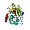
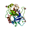
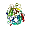
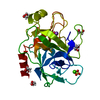
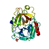
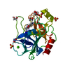
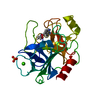
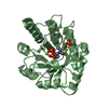
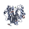
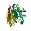
| Entry | Database: PDB / ID: 1h9h | ||||||
|---|---|---|---|---|---|---|---|
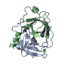
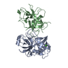
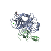
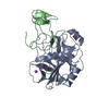
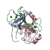
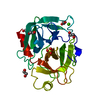
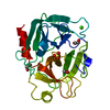
| Title | COMPLEX OF EETI-II WITH PORCINE TRYPSIN | ||||||
 Components Components |
| ||||||
 Keywords Keywords | HYDROLASE INHIBITOR / COMPLEX (SERINE PROTEASE-INHIBITOR) / TRYPSIN / SQUASH INHIBITOR / CYSTINE KNOT / HYDROLASE | ||||||
| Function / homology |  Function and homology information Function and homology informationtrypsin / digestion / serine-type endopeptidase inhibitor activity / serine-type endopeptidase activity / proteolysis / : / extracellular region / metal ion binding Similarity search - Function | ||||||
| Biological species |   ECBALLIUM ELATERIUM (jumping cucumber) ECBALLIUM ELATERIUM (jumping cucumber) | ||||||
| Method |  X-RAY DIFFRACTION / SYNCHROTRON / MOLECULAR REPLACEMENT / Resolution: 1.5 Å X-RAY DIFFRACTION / SYNCHROTRON / MOLECULAR REPLACEMENT / Resolution: 1.5 Å | ||||||
 Authors Authors | Kraetzner, R. / Wentzel, A. / Kolmar, H. / Uson, I. | ||||||
 Citation Citation | Journal: Acta Crystallogr.,Sect.D / Year: 2005 Title: Structure of Ecballium Elaterium Trypsin Inhibitor II (Eeti-II): A Rigid Molecular Scaffold Authors: Kraetzner, R. / Debreczeni, J.E. / Pape, T. / Schneider, T.R. / Wentzel, A. / Kolmar, H. / Sheldrick, G.M. / Uson, I. #1: Journal: J.Biol.Chem. / Year: 1999 Title: Sequence Requirements of the Gpng Beta-Turn of the Ecballium Elaterium Trypsin Inhibitor II Explored by Combinatorial Library Screening Authors: Wentzel, A. / Christmann, A. / Kraetzner, R. / Kolmar, H. #2: Journal: Biochemistry / Year: 1999 Title: Min-21 and Min-23, the Smallest Peptides that Fold Like a Cystine-Stabilized Beta-Sheet Motif: Design, Solution Structure, and Thermal Stability Authors: Heitz, A. / Le-Nguyen, D. / Chiche, L. #3: Journal: Proteins: Struct.,Funct., Genet. / Year: 1989Title: Use of Restrained Molecular Dynamics in Water to Determine Three-Dimensional Protein Structure: Prediction of the Three-Dimensional Structure of Ecballium Elaterium Trypsin Inhibitor II Authors: Chiche, L. / Gaboriaud, C. / Heitz, A. / Mornon, J.P. / Castro, B. / Kollman, P.A. | ||||||
| History |
| ||||||
| Remark 700 | SHEET DETERMINATION METHOD: DSSP THE SHEETS PRESENTED AS "B" IN EACH CHAIN ON SHEET RECORDS BELOW ... SHEET DETERMINATION METHOD: DSSP THE SHEETS PRESENTED AS "B" IN EACH CHAIN ON SHEET RECORDS BELOW IS ACTUALLY AN 6-STRANDED BARREL THIS IS REPRESENTED BY A 7-STRANDED SHEET IN WHICH THE FIRST AND LAST STRANDS ARE IDENTICAL. |
- Structure visualization
| Structure viewer | Molecule: MolmilJmol/JSmol |
|---|
- Downloads & links
-Download
| PDBx/mmCIF format | 1h9h.cif.gz | 67.2 KB | Display | PDBx/mmCIF format |
|---|---|---|---|---|
| PDB format | pdb1h9h.ent.gz | 49.1 KB | Display | PDB format |
| PDBx/mmJSON format | 1h9h.json.gz | Tree view | PDBx/mmJSON format | |
| Others |  Other downloads Other downloads |
-Validation report
| Arichive directory | https://data.pdbj.org/pub/pdb/validation_reports/h9/1h9hftp://data.pdbj.org/pub/pdb/validation_reports/h9/1h9h | HTTPS FTP |
|---|
-Related structure data
| Related structure data | 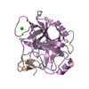 1h9iC  1w7zC  1ldtS S: Starting model for refinement C: citing same article ( |
|---|---|
| Similar structure data |


-Links
 PDBj
PDBj

- Assembly
| Deposited unit | 
| ||||||||
|---|---|---|---|---|---|---|---|---|---|
| 1 |
| ||||||||
| Unit cell |
|
-Components
| #1: Protein | Mass: 23493.496 Da / Num. of mol.: 1 / Source method: isolated from a natural source / Details: SIGMA / Source: (natural) |
|---|---|
| #2: Protein/peptide | Mass: 3902.500 Da / Num. of mol.: 1 / Mutation: YES / Source method: obtained synthetically / Details: C-TERMINAL TAG OF 6 HISTIDINES / Source: (synth.) ECBALLIUM ELATERIUM (jumping cucumber) / References: UniProt: P12071 |
| #3: Chemical | ChemComp-CA /   Mass: 40.078 Da / Num. of mol.: 1 / Source method: obtained synthetically / Formula: Ca Mass: 40.078 Da / Num. of mol.: 1 / Source method: obtained synthetically / Formula: Ca |
| #4: Water | ChemComp-HOH /  Mass: 18.015 Da / Num. of mol.: 177 / Source method: isolated from a natural source / Formula: H2O Mass: 18.015 Da / Num. of mol.: 177 / Source method: isolated from a natural source / Formula: H2O |
| Compound details | ENGINEERED MUTATION MET 7 ILE IN CHAIN I DUE TO RADIATION DAMAGE CYSTEINES WERE MODELLED PARTLY ...ENGINEERED |
| Has protein modification | Y |
| Sequence details | THE MICROHETEROGENEITY FOR THE CYSTEINE/SERINES AT RESIDUES 22,42,58, 136, 157, 191, 201, 220 ...THE MICROHETER |
-Experimental details
-Experiment
| Experiment | Method: X-RAY DIFFRACTION / Number of used crystals: 1 |
|---|
- Sample preparation
| Crystal | Density Matthews: 2.7 Å3/Da / Density % sol: 51 % |
|---|---|
| Crystal grow | pH: 6.7 / Details: pH 6.70 |
-Data collection
| Diffraction | Mean temperature: 100 K |
|---|---|
| Diffraction source | Source: SYNCHROTRON / Site: ESRF  / Beamline: ID14-2 / Wavelength: 0.9326 / Beamline: ID14-2 / Wavelength: 0.9326 |
| Detector | Type: MARRESEARCH / Detector: CCD / Date: Apr 4, 1999 / Details: TOROIDAL MIRROR |
| Radiation | Monochromator: DIAMOND (111), GE(220) / Protocol: SINGLE WAVELENGTH / Monochromatic (M) / Laue (L): M / Scattering type: x-ray |
| Radiation wavelength | Wavelength: 0.9326 Å / Relative weight: 1 |
| Reflection | Resolution: 1.5→20 Å / Num. obs: 49533 / % possible obs: 99.7 % / Redundancy: 8.05 % / Rmerge(I) obs: 0.0532 / Rsym value: 0.0278 / Net I/σ(I): 19.24 |
| Reflection shell | Resolution: 1.5→1.6 Å / Redundancy: 4.48 % / Rmerge(I) obs: 0.3755 / Mean I/σ(I) obs: 3.39 / Rsym value: 0.2779 / % possible all: 99.6 |
- Processing
| Software |
| |||||||||||||||||||||||||||||||||
|---|---|---|---|---|---|---|---|---|---|---|---|---|---|---|---|---|---|---|---|---|---|---|---|---|---|---|---|---|---|---|---|---|---|---|
| Refinement | Method to determine structure: MOLECULAR REPLACEMENT Starting model: PDB ENTRY 1LDT Resolution: 1.5→10 Å / Num. parameters: 8170 / Num. restraintsaints: 7778 / Cross valid method: FREE R-VALUE / σ(F): 0 / Stereochemistry target values: ENGH AND HUBER
| |||||||||||||||||||||||||||||||||
| Solvent computation | Solvent model: MOEWS & KRETSINGER, J.MOL.BIOL.91(1973)201-2 | |||||||||||||||||||||||||||||||||
| Refine analyze | Num. disordered residues: 17 / Occupancy sum hydrogen: 1746.9 / Occupancy sum non hydrogen: 1994.85 | |||||||||||||||||||||||||||||||||
| Refinement step | Cycle: LAST / Resolution: 1.5→10 Å
| |||||||||||||||||||||||||||||||||
| Refine LS restraints |
|
