 Movie
Movie Controller


+ Open data

- Basic information
| Entry | Database: PDB / ID: 1an1 | ||||||
|---|---|---|---|---|---|---|---|
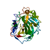
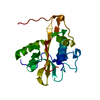
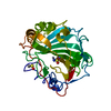
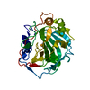
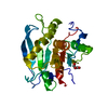
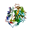
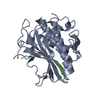
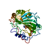
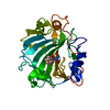
| Title | LEECH-DERIVED TRYPTASE INHIBITOR/TRYPSIN COMPLEX | ||||||
 Components Components |
| ||||||
 Keywords Keywords | COMPLEX (SERINE PROTEASE/INHIBITOR) / SERINE PROTEINASE INHIBITOR / TRYPTASE INHIBITION / NON-CLASSICAL KAZAL-TYPE INHIBITOR / COMPLEX (SERINE PROTEASE-INHIBITOR) / COMPLEX (SERINE PROTEASE-INHIBITOR) complex | ||||||
| Function / homology |  Function and homology information Function and homology informationtrypsin / digestion / serine-type endopeptidase inhibitor activity / serine-type endopeptidase activity / proteolysis / : / metal ion binding Similarity search - Function | ||||||
| Biological species |   Hirudo medicinalis (medicinal leech) Hirudo medicinalis (medicinal leech) | ||||||
| Method |  X-RAY DIFFRACTION / MOLECULAR REPLACEMENT / Resolution: 2.03 Å X-RAY DIFFRACTION / MOLECULAR REPLACEMENT / Resolution: 2.03 Å | ||||||
 Authors Authors | Priestle, J.P. / Di Marco, S. | ||||||
 Citation Citation | Journal: Structure / Year: 1997 Title: Structure of the complex of leech-derived tryptase inhibitor (LDTI) with trypsin and modeling of the LDTI-tryptase system. Authors: Di Marco, S. / Priestle, J.P. #1: Journal: Eur.J.Biochem. / Year: 1996Title: Purification, Characterization and Biological Evaluation of Recombinant Leech-Derived Tryptase Inhibitor (Rldti) Expressed at High Level in the Yeast Saccharomyces Cerevisiae Authors: Pohlig, G. / Fendrich, G. / Knecht, R. / Eder, B. / Piechottka, G. / Sommerhoff, C.P. / Heim, J. #2: Journal: Biol.Chem.Hoppe-Seyler / Year: 1994Title: A Kazal-Type Inhibitor of Human Mast Cell Tryptase: Isolation from the Medical Leech Hirudo Medicinalis, Characterization, and Sequence Analysis Authors: Sommerhoff, C.P. / Sollner, C. / Mentele, R. / Piechottka, G.P. / Auerswald, E.A. / Fritz, H. | ||||||
| History |
|
- Structure visualization
| Structure viewer | Molecule: MolmilJmol/JSmol |
|---|
- Downloads & links
-Download
| PDBx/mmCIF format | 1an1.cif.gz | 66.2 KB | Display | PDBx/mmCIF format |
|---|---|---|---|---|
| PDB format | pdb1an1.ent.gz | 47.7 KB | Display | PDB format |
| PDBx/mmJSON format | 1an1.json.gz | Tree view | PDBx/mmJSON format | |
| Others |  Other downloads Other downloads |
-Validation report
| Arichive directory | https://data.pdbj.org/pub/pdb/validation_reports/an/1an1ftp://data.pdbj.org/pub/pdb/validation_reports/an/1an1 | HTTPS FTP |
|---|
-Related structure data
| Related structure data | 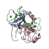 1eptS S: Starting model for refinement |
|---|---|
| Similar structure data |

-Links
 PDBj
PDBj

- Assembly
| Deposited unit | 
| ||||||||
|---|---|---|---|---|---|---|---|---|---|
| 1 |
| ||||||||
| Unit cell |
| ||||||||
| Components on special symmetry positions |
|
-Components
| #1: Protein | Mass: 23494.480 Da / Num. of mol.: 1 Source method: isolated from a genetically manipulated source Source: (gene. exp.) |
|---|---|
| #2: Protein/peptide | Mass: 4750.614 Da / Num. of mol.: 1 / Mutation: N115D (D-ASPARTIC ACID FORM) Source method: isolated from a genetically manipulated source Source: (gene. exp.) Hirudo medicinalis (medicinal leech) / Organ: SALIVARY GLANDS / Plasmid: PDP34 / Production host:  |
| #3: Chemical | ChemComp-CA /   Mass: 40.078 Da / Num. of mol.: 1 / Source method: obtained synthetically / Formula: Ca Mass: 40.078 Da / Num. of mol.: 1 / Source method: obtained synthetically / Formula: Ca |
| #4: Water | ChemComp-HOH /  Mass: 18.015 Da / Num. of mol.: 138 / Source method: isolated from a natural source / Formula: H2O Mass: 18.015 Da / Num. of mol.: 138 / Source method: isolated from a natural source / Formula: H2O |
| Has protein modification | Y |
-Experimental details
-Experiment
| Experiment | Method: X-RAY DIFFRACTION / Number of used crystals: 1 |
|---|
- Sample preparation
| Crystal | Density Matthews: 2.3 Å3/Da / Density % sol: 47 % | ||||||||||||||||||||
|---|---|---|---|---|---|---|---|---|---|---|---|---|---|---|---|---|---|---|---|---|---|
| Crystal grow | pH: 8.5 Details: 30% V/V 2-PROPANOL, 0.2M AMMONIUM ACETATE, 0.1M TRIS-HCL, PH 8.5 ROOM TEMPERATURE 27 MONTHS FOR GROWTH. Temp details: room temp | ||||||||||||||||||||
| Crystal | *PLUS | ||||||||||||||||||||
| Crystal grow | *PLUS Method: vapor diffusion, hanging drop | ||||||||||||||||||||
| Components of the solutions | *PLUS
|
-Data collection
| Diffraction | Mean temperature: 294 K |
|---|---|
| Diffraction source | Source: ROTATING ANODE / Type: ENRAF-NONIUS FR591 / Wavelength: 1.5418 |
| Detector | Type: MAR scanner 300 mm plate / Detector: IMAGE PLATE / Date: Jan 29, 1997 |
| Radiation | Monochromator: GRAPHITE(002) / Monochromatic (M) / Laue (L): M / Scattering type: x-ray |
| Radiation wavelength | Wavelength: 1.5418 Å / Relative weight: 1 |
| Reflection | Resolution: 2.03→29.14 Å / Num. obs: 17213 / % possible obs: 94.3 % / Observed criterion σ(I): 0 / Redundancy: 8.73 % / Rmerge(I) obs: 0.1154 / Rsym value: 0.1154 / Net I/σ(I): 27.82 |
| Reflection shell | Resolution: 2.03→2.1 Å / Redundancy: 6.2 % / Rmerge(I) obs: 0.4846 / Mean I/σ(I) obs: 4.7 / Rsym value: 0.4846 / % possible all: 79.4 |
| Reflection | *PLUS Num. measured all: 150264 |
| Reflection shell | *PLUS % possible obs: 79.4 % |
- Processing
| Software |
| ||||||||||||||||||||||||||||||||||||||||||||||||||||||||||||
|---|---|---|---|---|---|---|---|---|---|---|---|---|---|---|---|---|---|---|---|---|---|---|---|---|---|---|---|---|---|---|---|---|---|---|---|---|---|---|---|---|---|---|---|---|---|---|---|---|---|---|---|---|---|---|---|---|---|---|---|---|---|
| Refinement | Method to determine structure: MOLECULAR REPLACEMENT Starting model: PORCINE PANCREATIC BETA-TRYPSIN (PDB ENTRY 1EPT) Resolution: 2.03→6 Å / Rfactor Rfree error: 0.0076 / Data cutoff high absF: 1000000 / Data cutoff low absF: 0 / Isotropic thermal model: RESTRAINED / Cross valid method: THROUGHOUT / σ(F): 0 Details: RESIDUE 115 WAS REFINED AS D-ASPARTIC ACID. WATER MOLECULE 98 IS LOCATED ON A CRYSTALLOGRAPHIC 2-FOLD AXIS AND THEREFORE HAS AN OCCUPANCY OF 0.5
| ||||||||||||||||||||||||||||||||||||||||||||||||||||||||||||
| Displacement parameters | Biso mean: 26.5 Å2 | ||||||||||||||||||||||||||||||||||||||||||||||||||||||||||||
| Refinement step | Cycle: LAST / Resolution: 2.03→6 Å
| ||||||||||||||||||||||||||||||||||||||||||||||||||||||||||||
| Refine LS restraints |
| ||||||||||||||||||||||||||||||||||||||||||||||||||||||||||||
| LS refinement shell | Resolution: 2.03→2.09 Å / Rfactor Rfree error: 0.036 / Total num. of bins used: 12
| ||||||||||||||||||||||||||||||||||||||||||||||||||||||||||||
| Xplor file |
| ||||||||||||||||||||||||||||||||||||||||||||||||||||||||||||
| Software | *PLUS Name: X-PLOR / Version: 3.1 / Classification: refinement | ||||||||||||||||||||||||||||||||||||||||||||||||||||||||||||
| Refinement | *PLUS Rfactor obs: 0.17 / Rfactor Rwork: 0.17 | ||||||||||||||||||||||||||||||||||||||||||||||||||||||||||||
| Solvent computation | *PLUS | ||||||||||||||||||||||||||||||||||||||||||||||||||||||||||||
| Displacement parameters | *PLUS | ||||||||||||||||||||||||||||||||||||||||||||||||||||||||||||
| Refine LS restraints | *PLUS
|
